History: A 62-year-old woman presents with neck pain.
病史:62岁女性,颈部疼痛。
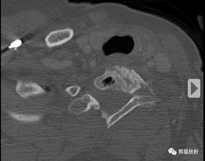
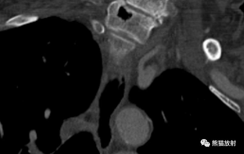
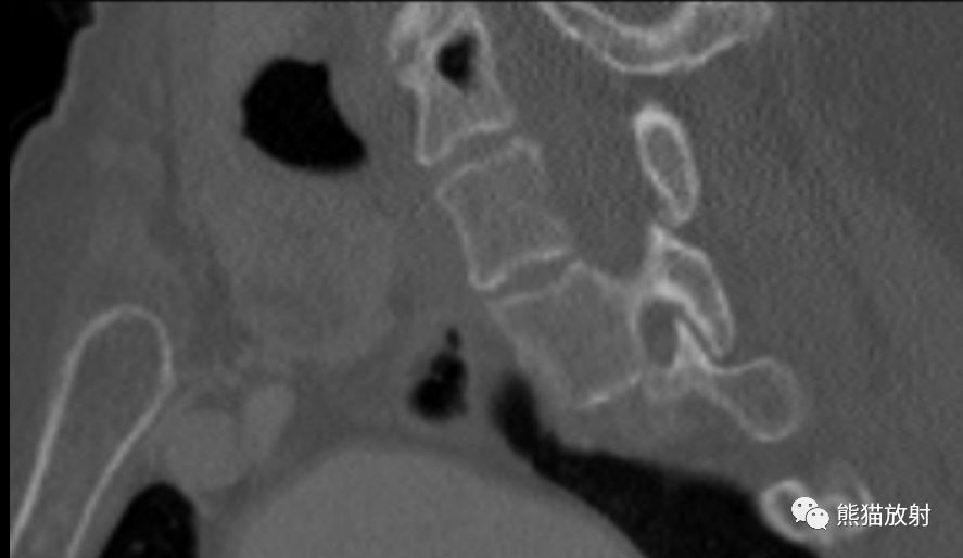
A CT scan was performed. Magnified axial, coronal, and sagittal images of the lower cervical spine are shown below.
行CT扫描,下颈椎的放大轴位、冠状、矢状图像如下所示。
Findings
CT demonstrates a focus of air within a lower cervical spine vertebral body that is completely contained within the vertebral body and does not appear to communicate with the disk space.
影像表现:
下颈椎椎体内见一局灶性气体影,它被完全包含在椎体内,不与椎间隙相交通。
Differential diagnosis
- Intravertebral pneumatocyst
- Schmorl\’s node
- Infection with gas-forming organism
鉴别诊断:
- 椎体内含气囊肿
- 许莫氏结节
- 产期微生物感染
Diagnosis: Intravertebral pneumatocyst
最后诊断:椎体内含气囊肿
Discussion
Intravertebral pneumatocyst
Intravertebral pneumatocysts are benign, gas-filled cystic lesions that occur within bone. Although they can occur in a variety of different locations, in the spine they most often occur in the cervical region. The etiology of these lesions is unknown, though their relation to degenerative change is likely.
- 良性,骨内,含气囊性病变。
- 尽管可见于不同部位,在椎体多见于颈椎。
- 病因不明,可能与退变有关。
Although these lesions were initially thought to be rare, subsequent studies have shown that they occur relatively frequently (present in up to 40% of patients undergoing cervical spine CT, with postsurgical and trauma patients excluded).
The lesions are considered benign, and no specific treatment is required.
- 最开始认为这些病变是罕见的,后来的研究发现该病比较常见,在颈椎CT检查的患者中高达40%,术后和外伤的患者除外。
- 为良性病变,无需特殊治疗。