History: A 62-year-old man presents with progressive ataxia and altered mental status for the past two months. He has a history of hepatitis C and hepatocellular carcinoma and is status post liver transplant.
病史:62岁男性,进行性共济失调及意识状态改变2个月。患有肝炎、肝癌,已行肝移植。
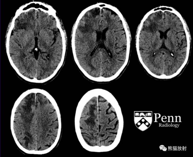
Axial images from a noncontrast head CT scan are shown in soft-tissue window below. 头颅CT平扫(轴位图像、软组织窗)
MR images
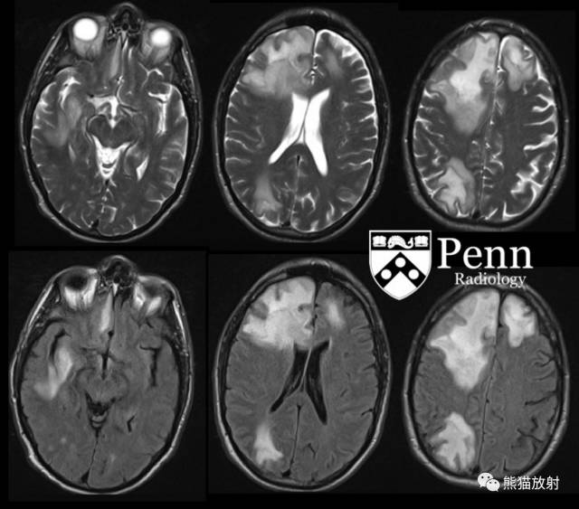
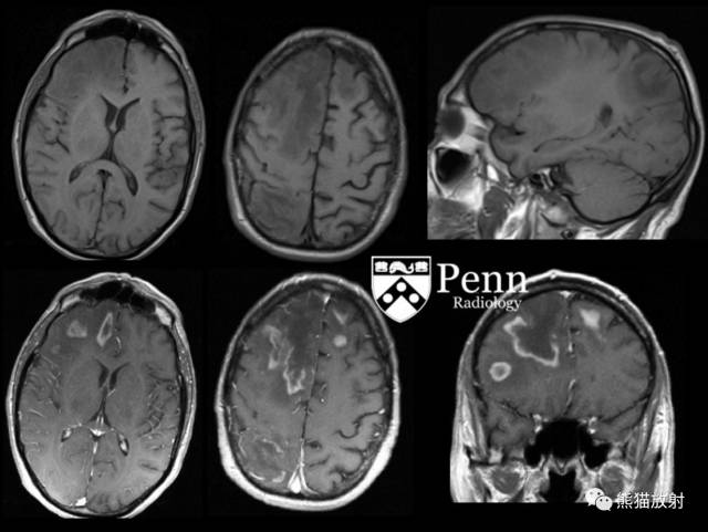
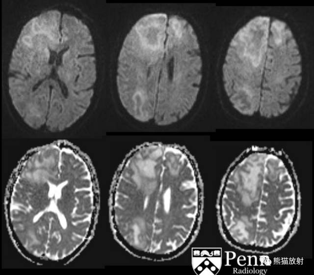
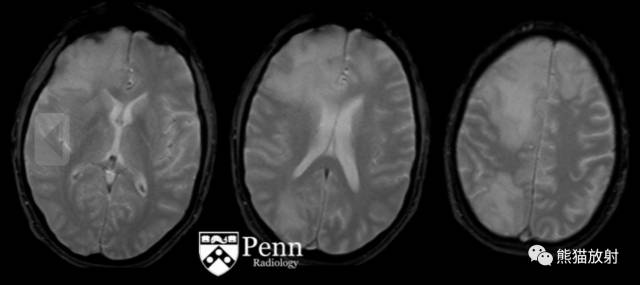
An MRI scan of the brain with and without contrast was obtained for further evaluation. Select axial T2-weighted, axial T2-weighted fluid-attenuated inversion-recovery (FLAIR), axial and sagittal T1-weighted, axial and coronal T1-weighted postcontrast, diffusion-weighted (DWI), apparent diffusion coefficient (ADC), and gradient-recalled echo (GRE) MR images of the brain are shown below.
进一步行颅脑MRI平扫 增强:轴位T2WI、轴位T2WI FLAIR、轴位及矢状T1WI、轴位及冠状增强、DWI、ADC图、GRE序列图像。
Findings
- Head CT:There are confluent regions of subcortical and deep white-matter hypoattenuation involving the bilateral frontal lobes, right more so than left, and right parietal lobe with mild effacement of the sulci. There are no findings of acute intracranial hemorrhage, extracerebral fluid collection, midline shift, or hydrocephalus.
- Brain MRI: There are multiple confluent regions of T2/FLAIR hyperintense signal abnormality predominantly involving the subcortical and deep white matter of the right greater than left frontal lobes and right parietal lobe corresponding to regions of hypoattenuation on CT. These areas demonstrate peripheral enhancement associated with a thin rim of restricted diffusion and surrounded by areas of milder T2 prolongation. There is relatively little associated mass effect. Additional small foci of enhancement are noted in the right occipital and temporal lobes. The large areas of confluent signal abnormality are most in keeping with demyelinating disease.
影像表现
CT:双侧额叶皮层下及深部白质融合性低密度灶,右侧为著,右顶叶脑沟轻度消失。未见急性颅内出血、脑外积水、中线异位或脑积水。
MRI:T2WI及FLAIR示双侧额叶皮层下及深部白质融合性(右侧为著)高信号灶,增强扫描边缘环形强化,相应区域弥散受限,周围可见轻度长T2信号区,占位效应相对较轻。另外,右枕叶及颞叶可见小强化灶。脑内大面积的融合性信号异常,最多见于脱髓鞘疾病。
Differential diagnosis
- Progressive multifocal leukoencephalopathy
- Acute demyelinating encephalitis
- Posterior reversible encephalopathy
- Tumefactive multiple sclerosis
- Limbic/autoimmune encephalitis
鉴别诊断:
- 进行性多灶性白质脑病
- 急性脱髓鞘性脑炎
- 脑后部可逆性脑病
- 肿瘤样多发性硬化
- 边缘系统/自身免疫性脑炎
Diagnosis: Progressive multifocal leukoencephalopathy
最后诊断:进行性多灶性白质脑病(PML)
PML是一种进行性亚急性脱髓鞘疾病,因 JC 病毒激活而导致局灶性或多灶性神经功能缺损,一般发生于细胞免疫反应缺陷患者,如AIDS,免疫抑制治疗后,淋巴增生性障碍。
病理:
- JC病毒在人类中广泛存在,潜伏于肾脏和脑,通常无症状。
- 在PML患者,JC病毒感染少突胶质细胞,导致髓鞘形成障碍或局部脱髓鞘。
- HIV感染患者一旦CD4计数小于100,发生PML的危险性增高。
- 经典PML:重度脱髓鞘并见肿胀的少突胶质细胞,缺乏或轻微的炎性改变。
- 炎性PML:可表现为广泛或局限性血管周围明显的炎性反应浸润,此外与经典PML相同。
影像表现:
- 皮层下及脑室旁白质,额顶叶为著,可见单发或多发融合不对称的密度减低区,FLAIR上呈高信号。
- DWI示病变外周可见片状弥散受限,其可能代表脱髓鞘或炎性反应的边缘。
- 经典PML:无明显强化或占位效应;
- 炎性PML:外周环形强化,有或无占位效应及血管源性水肿。
诊断:神经影像学结合脑脊液聚合酶链反应(PCR)证实了 JC 病毒 DNA。
治疗:尚无针对JC病毒的特异性治疗方法。对于HIV感染患者,高效抗逆转录病毒疗法(HAART)为最佳选择,生存期从过去的2至6个月,现可改善为3至4年。