History: A 33-year-old woman presents with anterior knee pain with weight bearing and give-away sensation. Symptoms did not improve with joint steroid injection.
病史:33岁女性,负重时或无意中膝前部疼痛,关节内注射类固醇后症状无缓解。
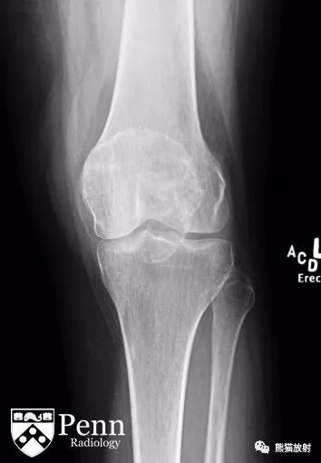
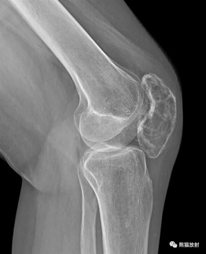
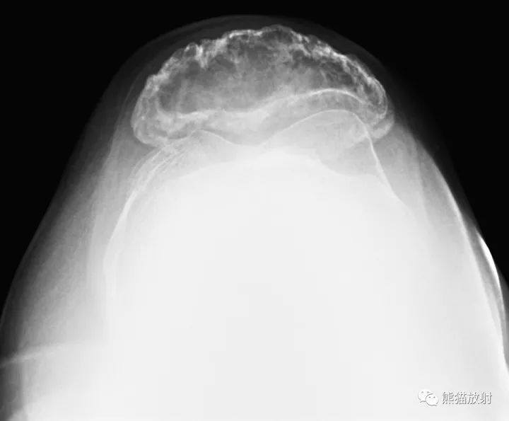
Frontal, lateral, and patellar radiographs of the left knee are shown below.
左膝正位、侧位、髌骨位如下所示。
MR images
Findings
Radiographs: Radiographs demonstrate an expansile lytic process involving the entire patella with numerous septations and lobulation of the cortical margins. The visualized portions of the femur, tibia, and fibula appear normal. No joint effusion is demonstrated.
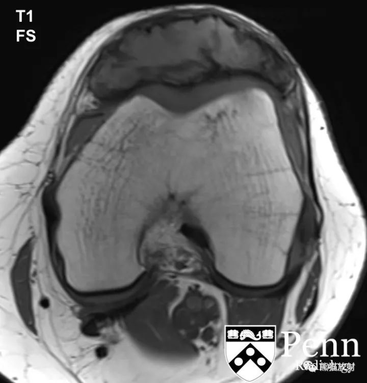
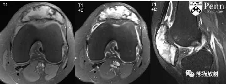
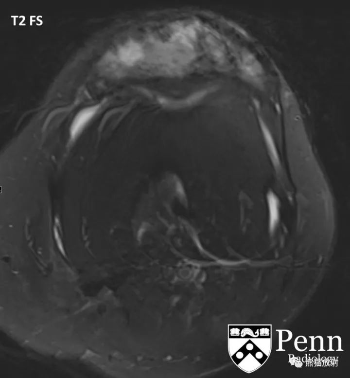
MRI: MR images demonstrates an expansile lesion centered and replacing the patella with associated cortical thinning. The mass demonstrates predominantly T1 iso- to hyperintense and heterogeneous T2 hyperintense signal with some areas of relative T2 hypointense signal and associated heterogeneous postcontrast enhancement.
影像表现:
平片:整个髌骨膨胀性溶骨性病变,内见大量分隔,骨皮质边缘呈分叶状。所见股骨、胫骨、腓骨未见异常,无关节积液。
MRI:髌骨膨胀性病变,皮质变薄,病灶在T1WI上呈等、稍高信号,T2WI上呈不均质高信号,局部见低信号,增强扫描不均质强化。
Differential diagnosis
- Giant cell tumor
- Aneurysmal bone cyst
- Expansile lytic metastatic disease
- Multiple myeloma
- Chondroblastoma
- Telangiectatic osteosarcoma
- Giant cell-rich osteosarcoma
鉴别诊断:
- 骨巨细胞瘤
- 动脉瘤样骨囊肿
- 膨胀性溶骨性转移性病变
- 多发性骨髓瘤
- 软骨母细胞瘤
- 毛细血管扩张型骨肉瘤
- 富巨细胞型骨肉瘤
Diagnosis: Giant cell tumor in the patella
最后诊断:(髌骨)骨巨细胞瘤
Key points
Giant cell tumor (GCT)
Epidemiology
- Primary tumors of the patella are extremely rare; only case reports are available. The most common tumors of the patella include giant cell tumors and chrondroblastomas.
- Giant cell tumors usually occur in patients between ages 20 and 50 and peak during the third decade.
流行病学
髌骨原发性肿瘤极其罕见,仅见个案报道,髌骨最常见的肿瘤包括骨巨细胞瘤及软骨母细胞瘤;
GCT多见于20-50岁患者,发病高峰在三十多岁。
Pathophysiology
- These tumors are composed of mononuclear stromal cells and multinucleated giant cells that exhibit osteoclastic activity.
- The stromal cells have overexpression of receptor activator of nuclear factor kappa-B ligand (RANKL) that promote recruitment of numerous reactive multinucleated osteoclast-like giant cells. This causes lacunar bone resorption.
- Giant cell tumors usually involve long bones (75% to 90%), particularly the knee, but they can occur in any bone.
- They are benign tumors, but they can be locally aggressive and recur in the surgical bed.
- Lung metastasis may occur in 1% to 6% of the cases that are histologically benign. It is usually related to trauma and hematogenous seeding.
病理生理学
肿瘤由单核基质细胞及多核巨细胞组成,并显示破骨活动。
基质细胞的受体激动剂核因子kappa-B配体(RANKL)的过渡表达,促进大量活性多核破骨样巨细胞形成。这导致多灶性骨吸收。
GCT常累及长骨 (75%-90%),尤其是膝部,但其实GCT可见于任何骨。
良性肿瘤,但可局部侵袭或术后复发。
1%-6%的病例可见肺转移,在组织学上为良性,通常与外伤及血行种植相关。
Clinical presentation
- Patients may have nonspecific pain.
- Giant cell tumors can be found as incidental findings.
临床表现:
患者可有非特异性疼痛;
GCT可为偶然发现。
Imaging features
Radiographs:
- Occurs in patients with closed physes.
- Extends near the articular surface/subchondral bone.
- Eccentric in location.
- Appears as a lesion with a well-defined but nonsclerotic margin.
- Can have aggressive features, such as cortical expansion and cortical destruction with or without a soft-tissue component.
- Can be complicated by pathologic fractures.
- Fluid-fluid levels are suggestive of a secondary formation of aneurysmal bone cysts, which occur in 14% of the cases.
- After surgical resection, recurrence is suggested by the development of progressive lucency at the cement-bone interface.
MRI:
- Giant cell tumors have a nonspecific appearance on MRI.
- On T1-weighted sequences, they have intermediate to decrease signal intensity.
- With fluid-sensitive sequences, they have high signal intensity.
- They show enhancement after contrast administration.
Nuclear medicine: Bone scan with methylene diphosphonate (MDP) shows increased peripheral radiotracer uptake in giant cell tumors. The center is photopenic due to central necrosis or osteolysis.
影像表现:
平片:
- 发生在骨骺闭合患者;
- 病灶可达关节面下或软骨下骨;
- 位置为偏心性;
- 边界清楚,但无硬化边;
- 可具有侵袭性表现,如:骨皮质膨胀或破坏,有或无软组织肿块;
- 可合并病理性骨折;
- 14%的病例可继发动脉瘤样骨囊肿形成,并见液液平面;
- 手术切除后,在骨水泥-骨交界面出现进行性透亮区,提示肿瘤复发。
MRI:
- GCT在MRI上的表现不具有特异性;
- T1WI上呈中等或较低信号;
- 在对水更敏感的序列上呈高信号;
- 增强后可见强化。
核医学:MDP骨扫描示GCT周边摄取增高,中心摄取较低(由于坏死或骨质溶解)。