

Findings
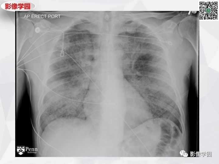
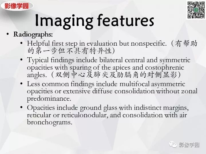
- Chest radiograph: Diffuse bilateral ill-defined airspace opacities are seen in a variable distribution.
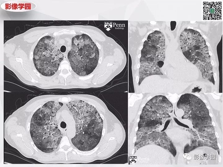
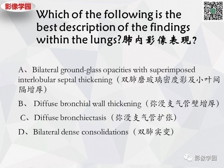
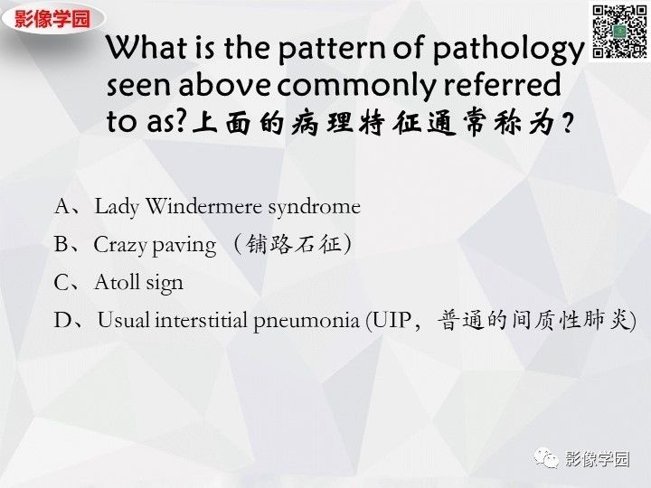
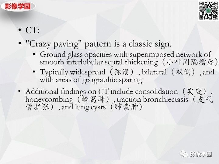
- Chest CT: CT demonstrates diffuse bilateral ground-glass opacities with superimposed interlobular septal thickening in a “crazy paving” pattern with regions of geographic sparing.
Differential diagnosis
- Pulmonary alveolar proteinosis
- Acute respiratory distress syndrome (ARDS)
- Severe pulmonary edema
- Diffuse alveolar hemorrhage
- Pneumocystis jirovecii infection
Diagnosis: Pulmonary alveolar proteinosis
The patient underwent bronchoalveolar lavage, which demonstrated milky and turbid fluid with thick sediment. Cytology demonstrated large foamy macrophages. Serum antigranulocyte-macrophage colony-stimulating factor (anti-GM-CSF) autoantibodies were positive.