History: Boy presents with knee pain.
病史:男孩,膝部疼痛。
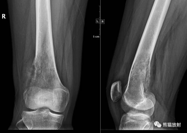
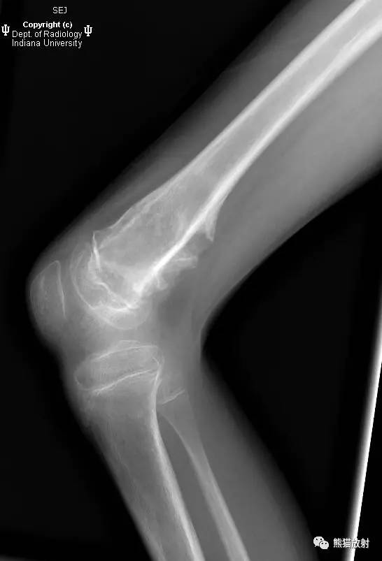
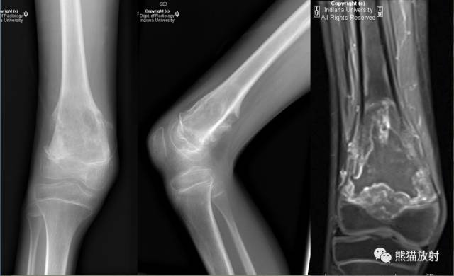
Radiographs are shown below. 平片如下所示。
MR images to correlate with the radiographs are shown below.
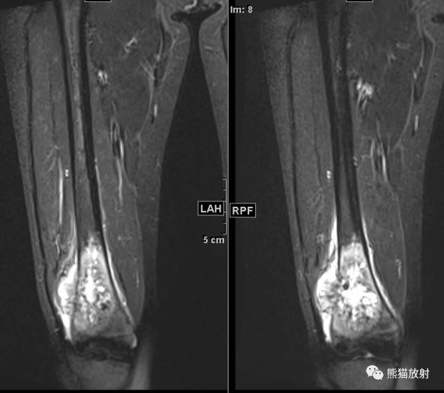
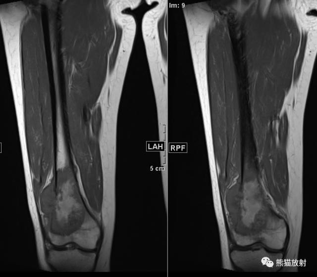
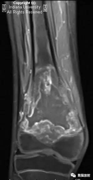
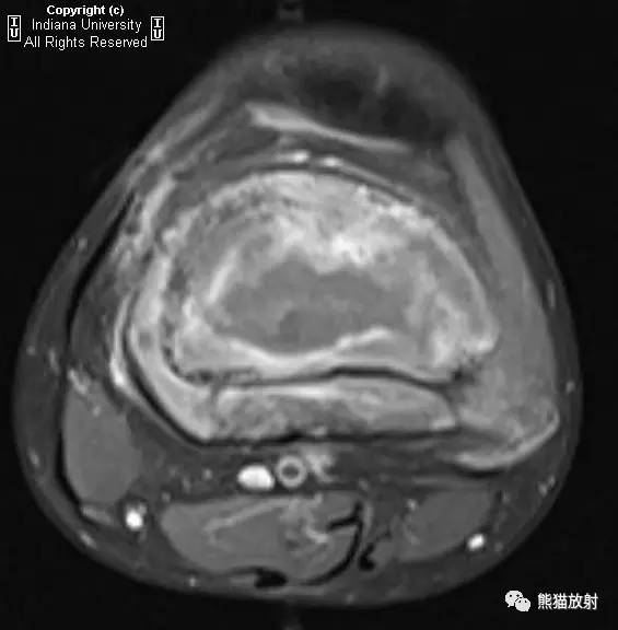
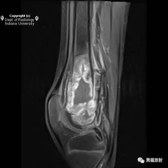
与平片相对应的MR图像如下所示。
Findings
There is a destructive lesion within the distal femoral metaphysis with elevation of the periosteum and an associated soft-tissue lesion without matrix.
影像表现:
股骨远侧干骺端可见骨质破坏区,骨膜反应,软组织肿块,未见骨基质。
Differential diagnosis
- Osteosarcoma
- Aneurysmal bone cyst
- Osteomyelitis
- Lymphoma
鉴别诊断:
- 骨肉瘤
- 动脉瘤样骨囊肿
- 骨髓炎
- 淋巴瘤
Discussion
Osteosarcoma
Osteosarcoma is an aggressive, destructive primary neoplasm of the bone. It is a malignant tumor that can produce osteoid matrix and, hence, often presents itself during periods of rapid bone growth, such as childhood and adolescence. A second incidence peak also occurs after age 60.
骨肉瘤是一种具有侵袭性、破坏性的原发性骨肿瘤,是一种能产生骨样基质的恶性肿瘤,因此,肿瘤常发生在骨生长迅速的时期,例如儿童或青少年,第二个发病高峰则在60岁之后。
Patients present with progressive pain and swelling at the affected site. The pain often worsens at night and is not relieved with rest. It most commonly affects the metaphysis of long bones and can extend into the epiphysis. The knee is the most common site affected.
患者表现为病变部位进行性疼痛和肿胀,疼痛常于夜晚加重,休息后无缓解。好发于长骨干骺端,可累及至骨骺。膝关节为最常见的发病部位。
Due to its aggressive nature, surgical resection with chemotherapy is the conventional treatment option. The five-year survival is 41%, although it increases to 70% if there is no metastasis.
由于肿瘤侵袭性生长,手术切除并化疗为常用的治疗手段,五年生存率为41%,如果没有转移可增加至70%。
Radiologic overview of the diagnosis
Radiography is the usual initial study and shows a destructive intramedullary lesion with extension to the bony cortex. An associated soft-tissue mass also can be seen.There will be a wide zone of transition and periosteal reaction. Fluid-fluid levels similar to that seen in an aneurysmal bone cyst also may be seen. When these findings are seen on radiography, MRI is usually the next study of choice.
MRI shows a destructive lesion that enhances with contrast. The soft-tissue component will demonstrate increased T2 signal. The bone will demonstrate intermediate T1 signal, greater than that of normal bone. When imaging, the entire bone should be imaged instead of just the area of interest, as skip lesions are not uncommon.
影像诊断概述
平片是最常用的首要检查方法,表现为髓内破坏性病变,扩大至骨皮质,并可见软组织肿块。可见较大范围的过渡区及骨膜反应。偶尔可见类似动脉瘤样骨囊肿的液液平面,有此类发现时,需进一步行MRI扫描。
MRI表现为骨质破坏病变,增强有强化,软组织成分表现为长T2信号,病变骨呈等T1WI信号,信号高于正常骨质。由于跳跃病灶较为常见,成像时需完整显示整个骨骼,而不是只扫描原有病变区域。
Key points
- Osteosarcoma is an aggressive primary neoplasm of the bone.
- Osteosarcoma has two peak incidences: one during childhood and adolescence and one after age 60.
- The most common presenting symptom is pain, which is most severe at night.
- MRI is the study of choice to evaluate osteosarcoma. Skip lesions are common, so the entire bone should be imaged.
知识点
- 骨肉瘤是一种具有侵袭性的原发性骨肿瘤;
- 两个发病高峰:儿童及青少年、60岁以后;
- 常见临床表现为疼痛,夜间加重;
- 最好选择MRI评估骨肉瘤,因跳跃病灶多见,需将发病的骨骼完整成像。
青少年、长骨干骺端、骨质破坏、骨膜反应、软组织肿块、明显不均质强化。
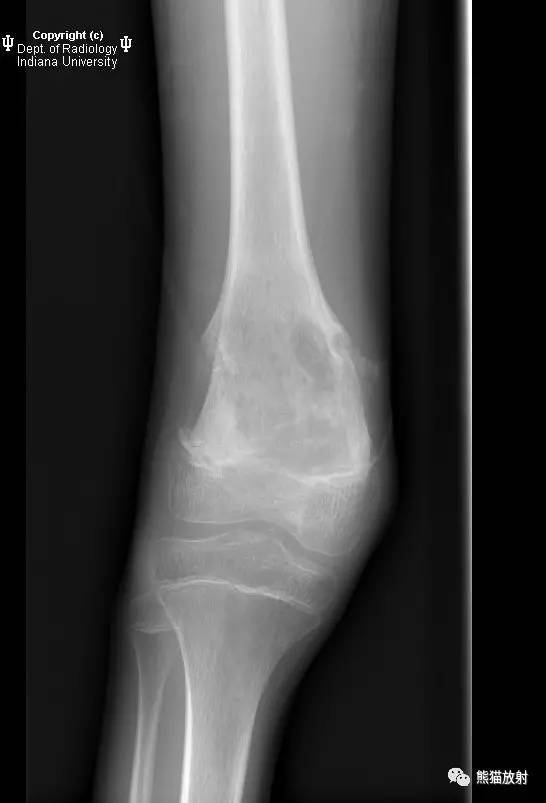
补充病例:
女,20岁,右膝疼痛1月余。
(右股骨远端)普通型骨肉瘤
X线平片正侧位、MR 冠状T2WI压脂、冠状T1WI