
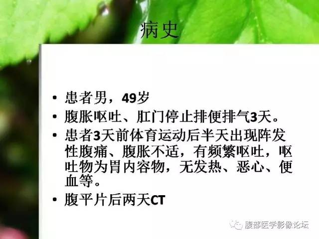
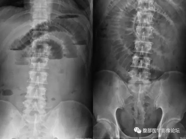
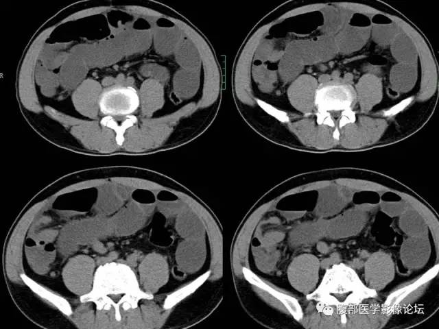
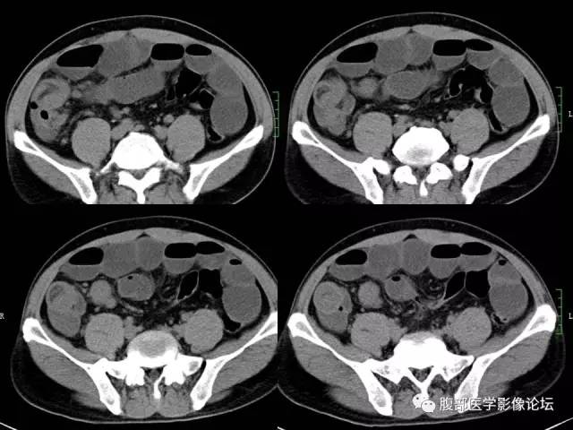
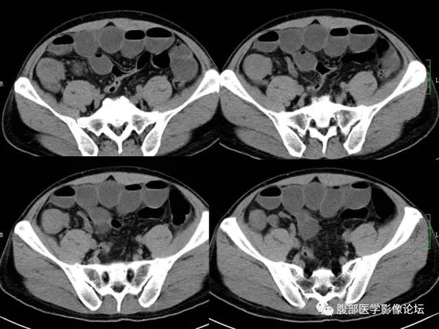
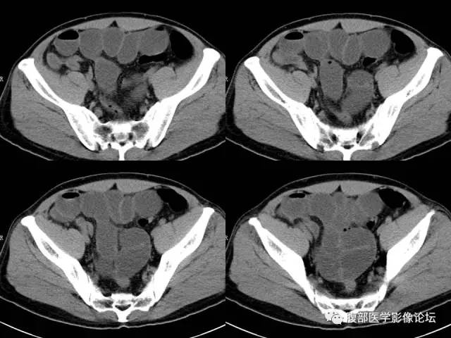
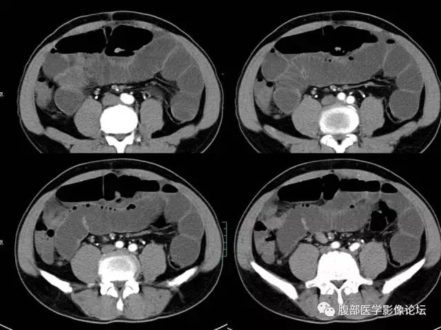
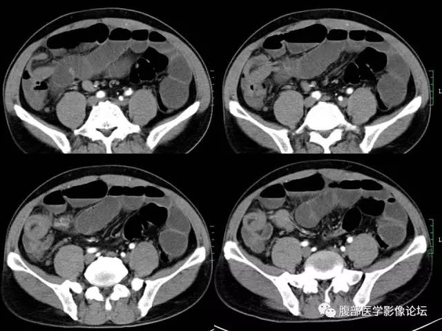
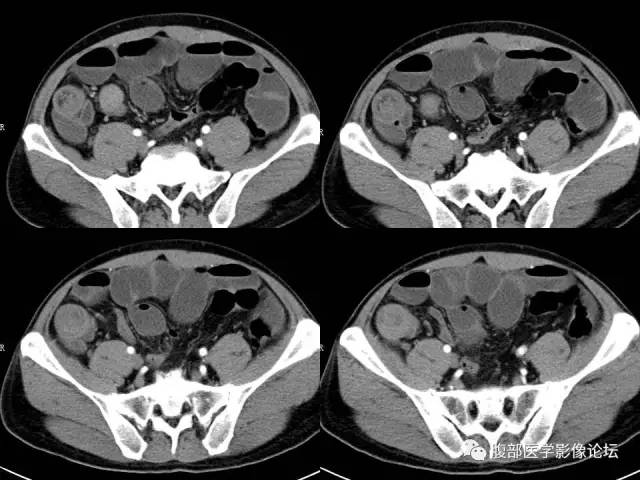
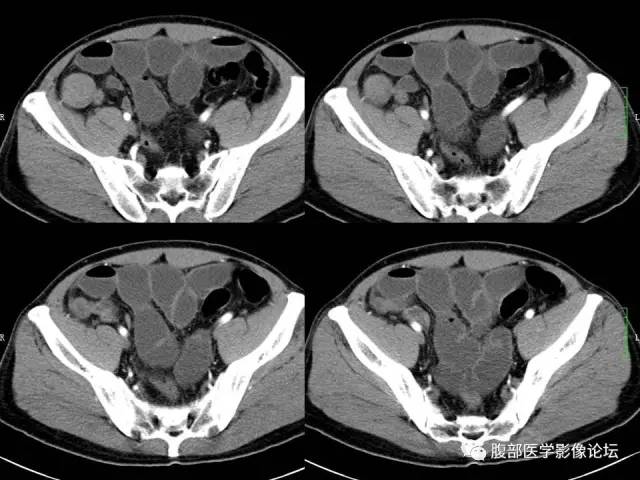
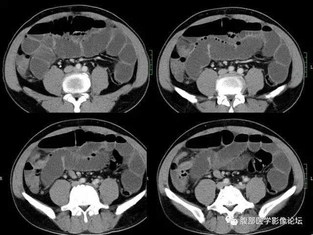
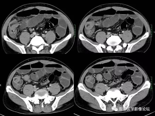
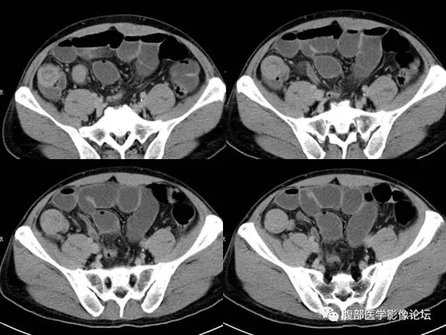
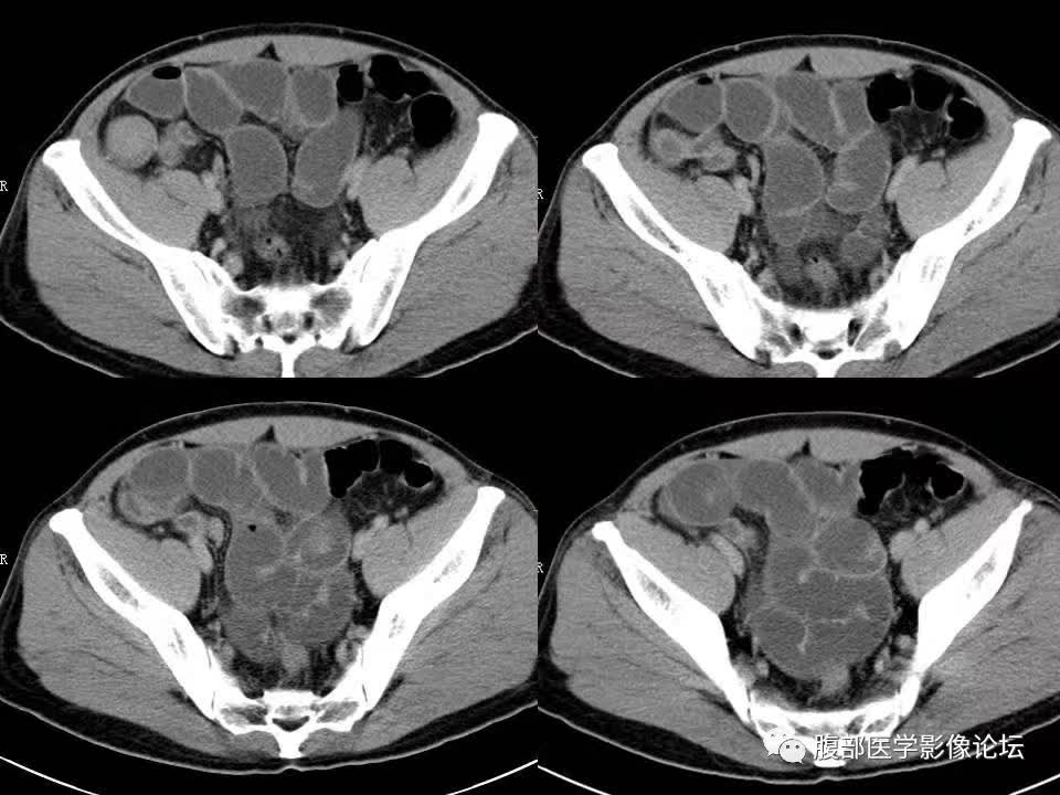
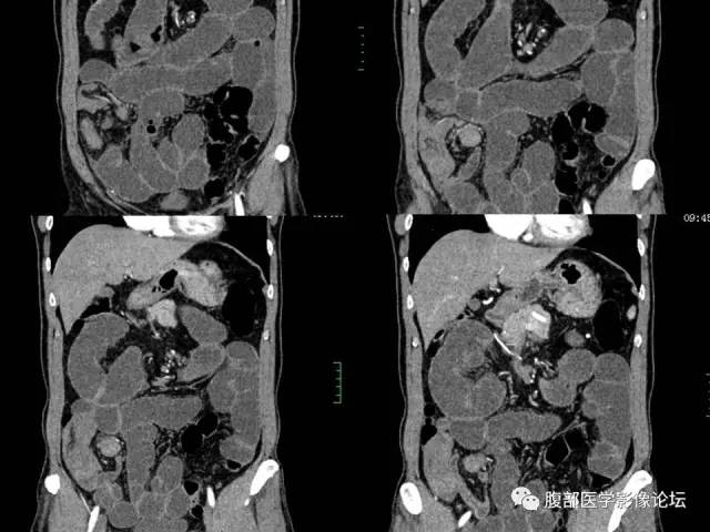
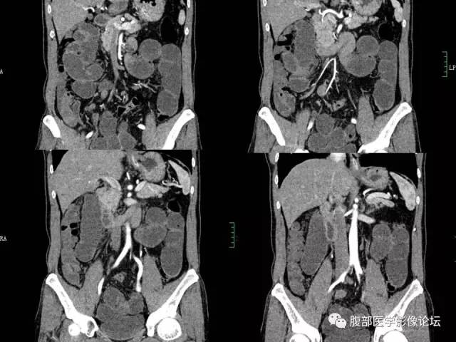
【病例】回肠淋巴瘤并肠套叠1例CT

17 2 月

【病例】回肠淋巴瘤并肠套叠1例CT

【病例】回肠淋巴瘤并肠套叠1例CT

【病例】回肠淋巴瘤并肠套叠1例CT

【病例】回肠淋巴瘤并肠套叠1例CT

【病例】回肠淋巴瘤并肠套叠1例CT

【病例】回肠淋巴瘤并肠套叠1例CT

【病例】回肠淋巴瘤并肠套叠1例CT

【病例】回肠淋巴瘤并肠套叠1例CT

【病例】回肠淋巴瘤并肠套叠1例CT

【病例】回肠淋巴瘤并肠套叠1例CT

【病例】回肠淋巴瘤并肠套叠1例CT

【病例】回肠淋巴瘤并肠套叠1例CT

【病例】回肠淋巴瘤并肠套叠1例CT

【病例】回肠淋巴瘤并肠套叠1例CT

【病例】回肠淋巴瘤并肠套叠1例CT

【病例】回肠淋巴瘤并肠套叠1例CT

【病例】回肠淋巴瘤并肠套叠1例CT

【病例】回肠淋巴瘤并肠套叠1例CT

【病例】回肠淋巴瘤并肠套叠1例CT

【病例】回肠淋巴瘤并肠套叠1例CT

【病例】回肠淋巴瘤并肠套叠1例CT

【病例】回肠淋巴瘤并肠套叠1例CT

【病例】回肠淋巴瘤并肠套叠1例CT

【病例】回肠淋巴瘤并肠套叠1例CT

【病例】回肠淋巴瘤并肠套叠1例CT

【病例】回肠淋巴瘤并肠套叠1例CT

【病例】回肠淋巴瘤并肠套叠1例CT

病理

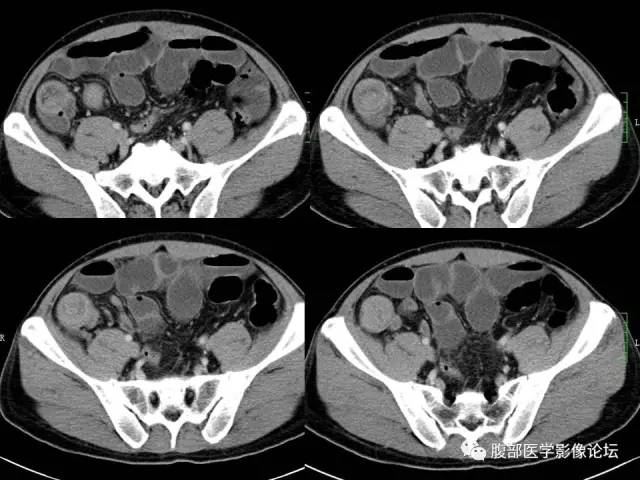
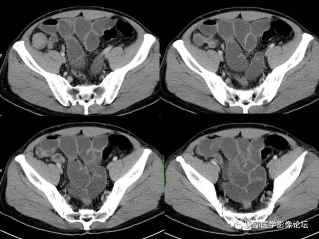
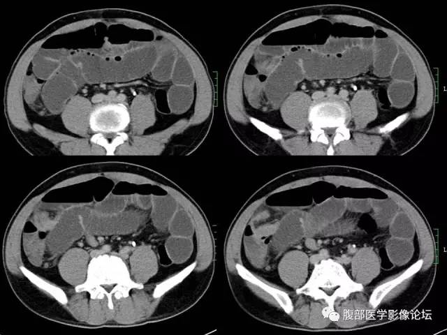
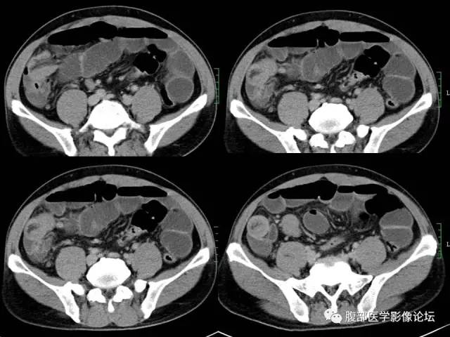
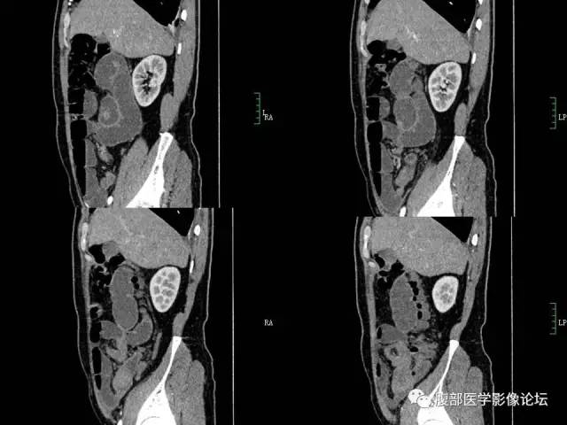
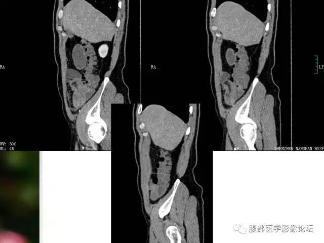
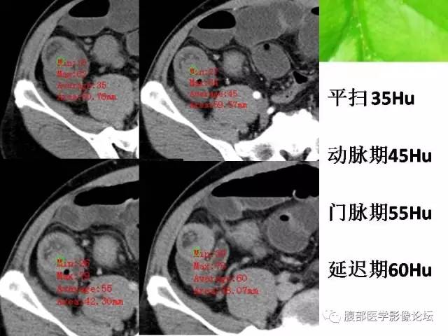
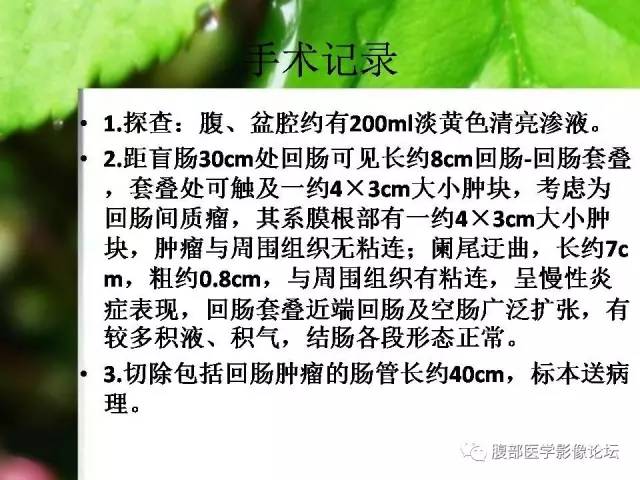
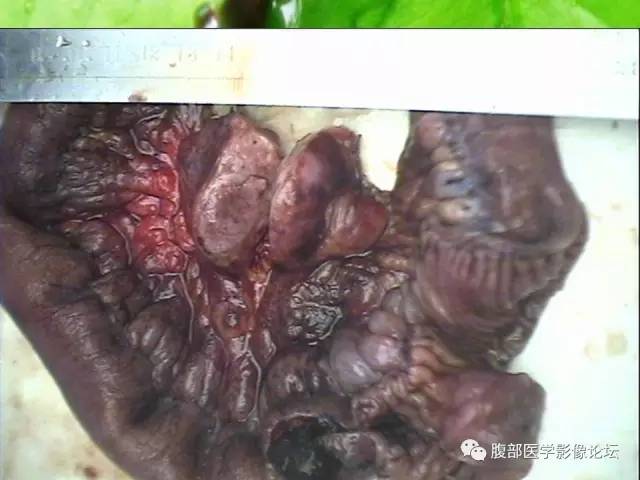
1、(小肠及肠系膜肿物)肠管一段,长40cm,直径2.5cm,距一侧断端12cm处见管腔内见一4×3×2cm的肿物,切面灰白、灰红,另见肠系膜一肿物4×3×1.5cm,切面灰红,质中。

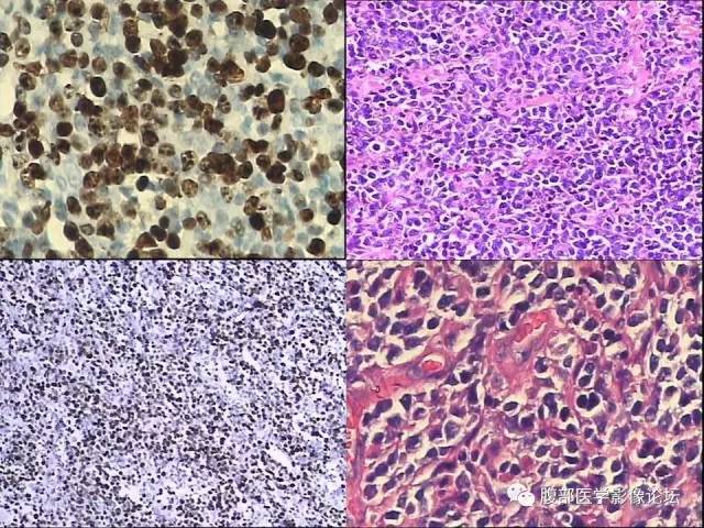
2、镜下正常肠壁结构消失,间质血管增生,代之以弥漫性成片增生的体积较大的肿瘤细胞,部分瘤细胞可见核仁,核分裂像多见,瘤细胞胞浆丰富、略嗜酸性。免疫组化结果:CD45( )、CD20( )、CD79α( )、CD10( )、CD5( )、MUM-1( )、Vimentin( )、CD45RO(部分阳性)、CD3(部分阳性)、BCL-6局灶( )、BCL-2(-)、CD30(-)、CD56(-)、Cyclin D1(-)、MPO(-)、CK(-)、Ki67约80%肿瘤细胞阳性。

(回肠及回肠系膜肿物)结合免疫组化结果,符合弥漫性大B细胞淋巴瘤,肠管两端切缘未见肿瘤累及。

发表回复

您的邮箱地址不会被公开。 必填项已用 * 标注