
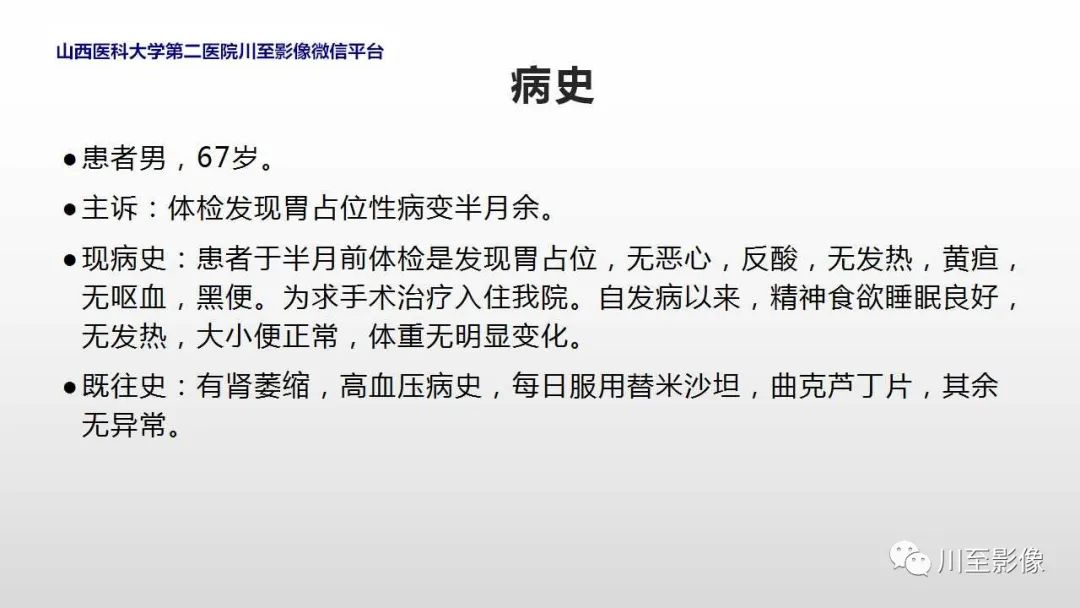
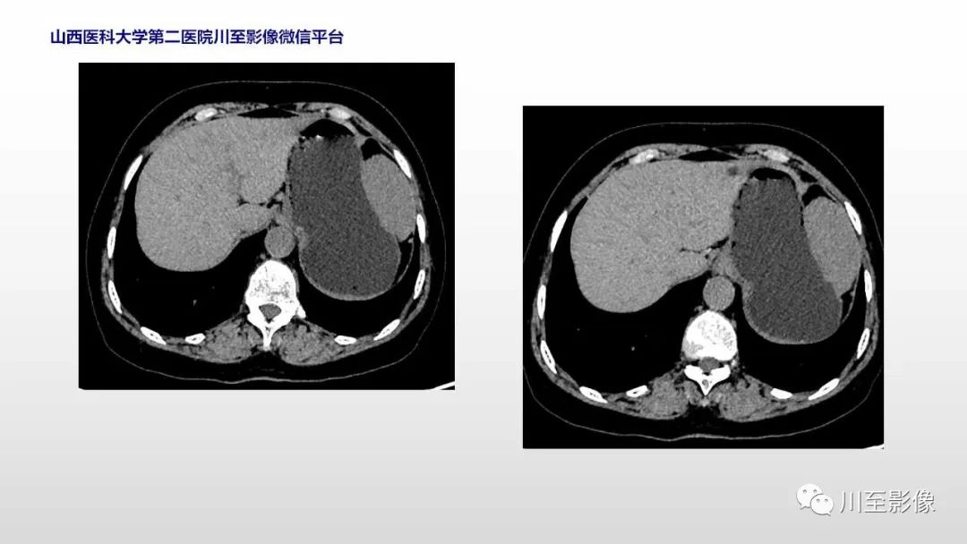
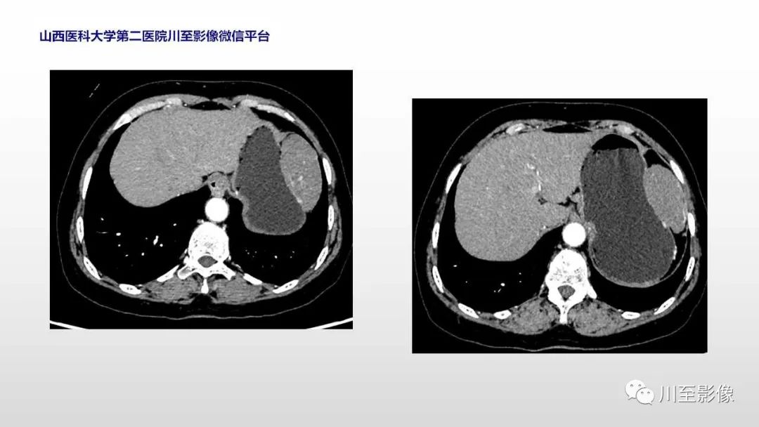
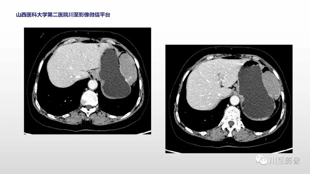
【PPT】胃肠道间质瘤

17 8 月

影像PPT - 【PPT】胃肠道间质瘤-1

影像PPT - 【PPT】胃肠道间质瘤-2

影像PPT - 【PPT】胃肠道间质瘤-3

影像PPT - 【PPT】胃肠道间质瘤-4

影像PPT - 【PPT】胃肠道间质瘤-5

影像PPT - 【PPT】胃肠道间质瘤-6

影像PPT - 【PPT】胃肠道间质瘤-7

影像PPT - 【PPT】胃肠道间质瘤-8

影像PPT - 【PPT】胃肠道间质瘤-9

影像PPT - 【PPT】胃肠道间质瘤-10

影像PPT - 【PPT】胃肠道间质瘤-11

影像PPT - 【PPT】胃肠道间质瘤-12

影像PPT - 【PPT】胃肠道间质瘤-13

影像PPT - 【PPT】胃肠道间质瘤-14

影像PPT - 【PPT】胃肠道间质瘤-15

影像PPT - 【PPT】胃肠道间质瘤-16

影像PPT - 【PPT】胃肠道间质瘤-17

影像PPT - 【PPT】胃肠道间质瘤-18

影像PPT - 【PPT】胃肠道间质瘤-19

影像PPT - 【PPT】胃肠道间质瘤-20

影像PPT - 【PPT】胃肠道间质瘤-21

影像PPT - 【PPT】胃肠道间质瘤-22

影像PPT - 【PPT】胃肠道间质瘤-23

影像PPT - 【PPT】胃肠道间质瘤-24

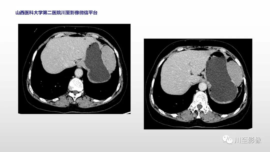
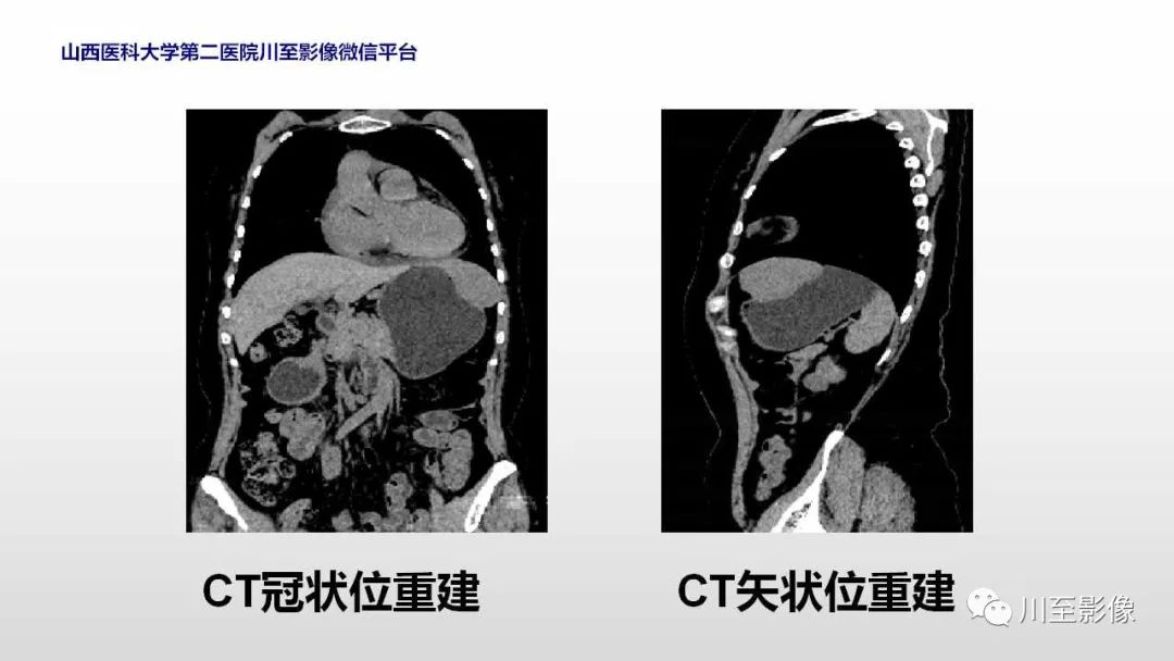
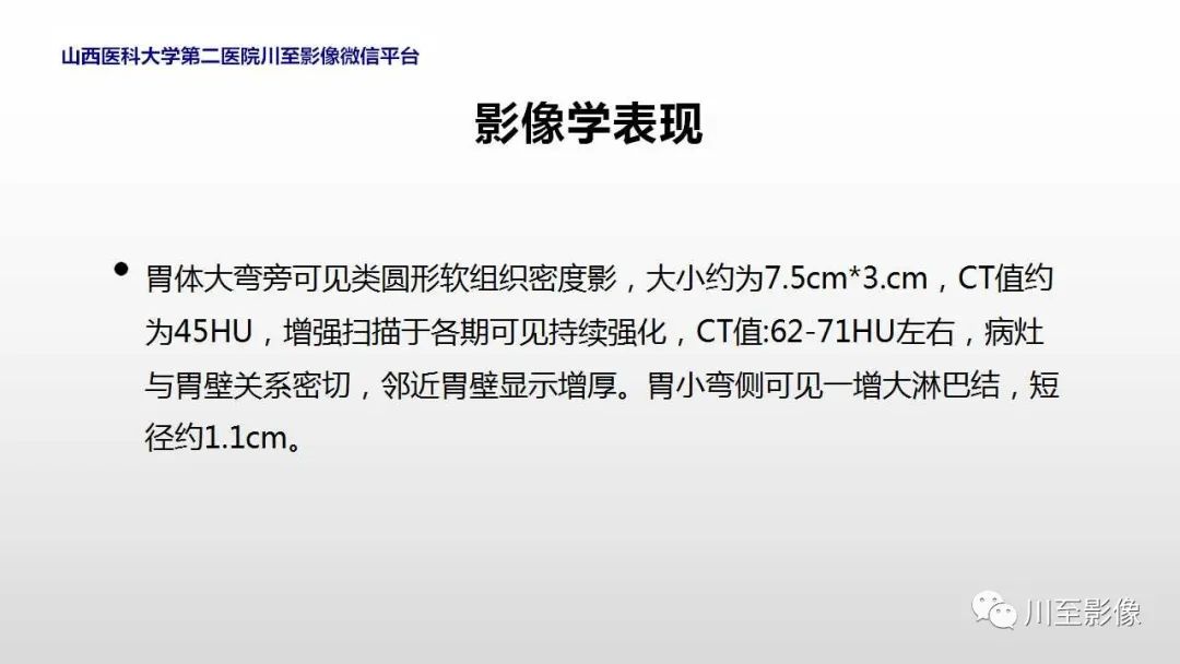
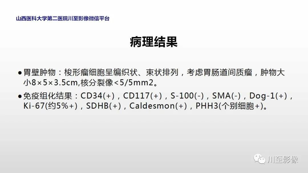
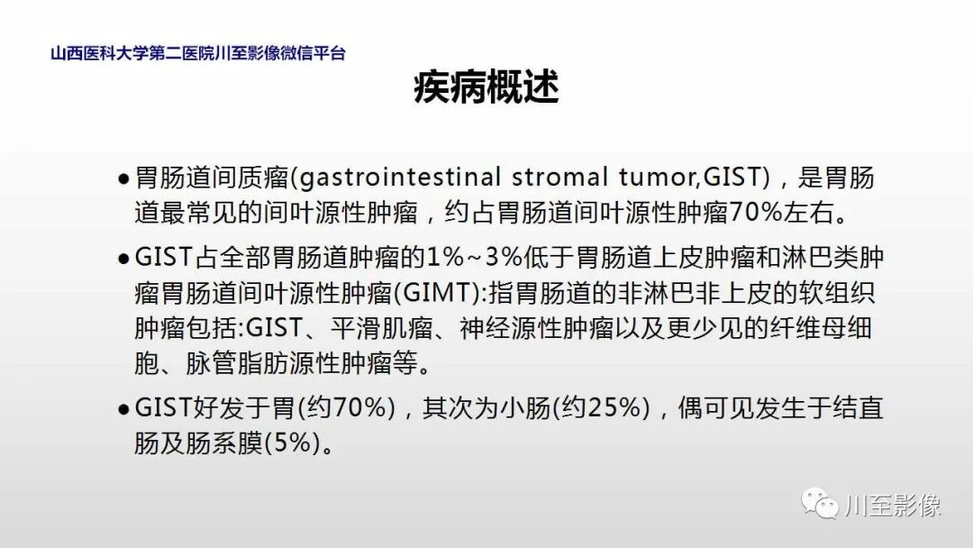
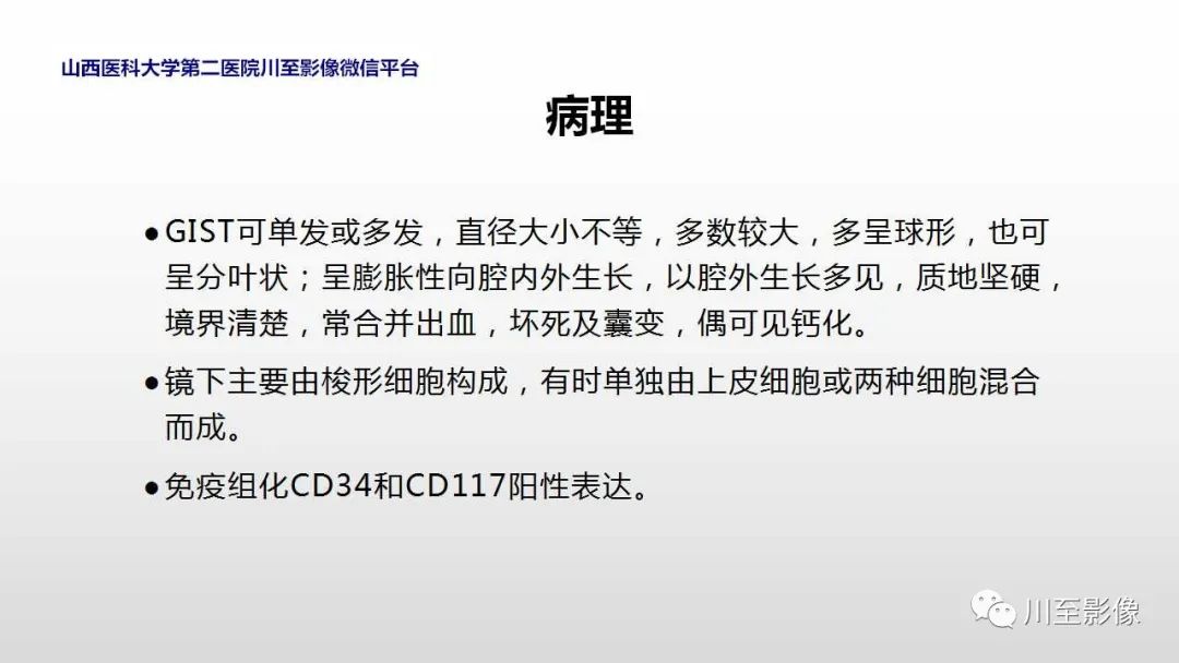
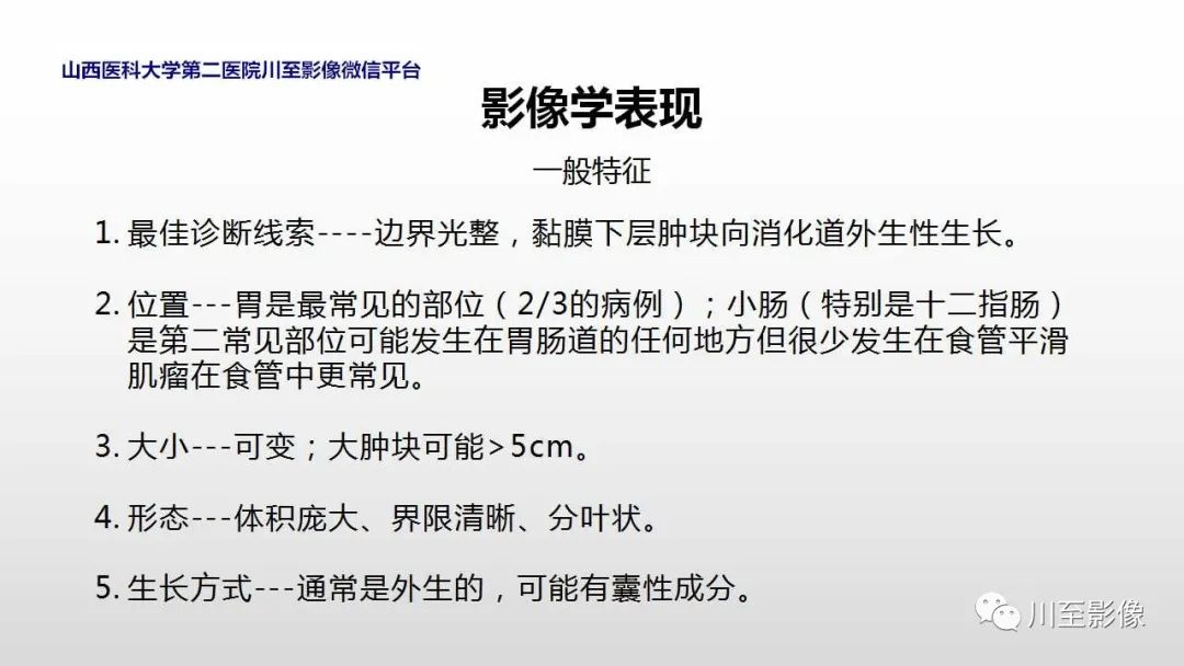
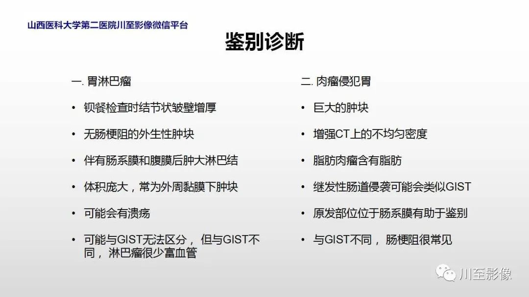
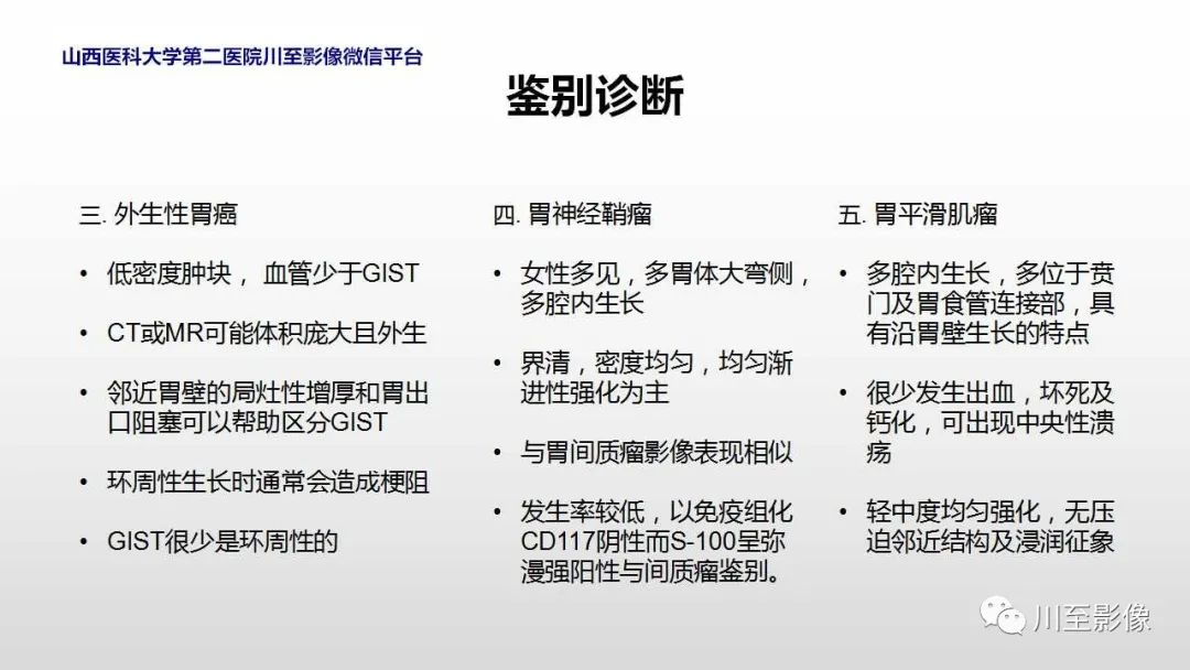
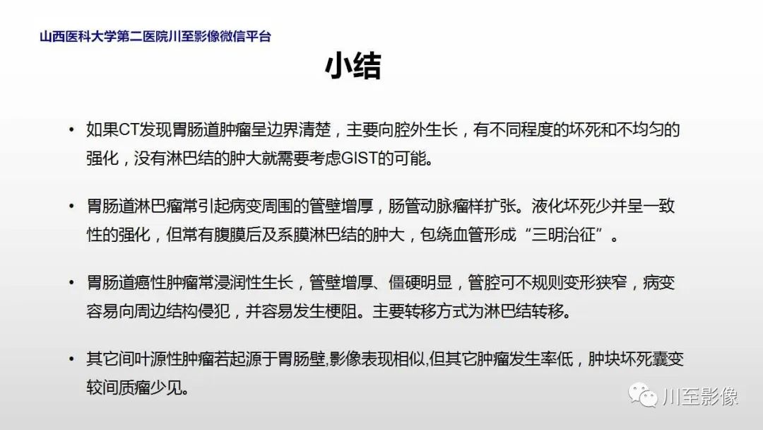
胃肠道间质瘤(gastrointestinal stromal tumor,GIST),是胃肠道最常见的间叶源性肿瘤,约占胃肠道间叶源性肿瘤70%左右。GIST占全部胃肠道肿瘤的1%~3%低于胃肠道上皮肿瘤和淋巴类肿瘤胃肠道间叶源性肿瘤(GIMT):指胃肠道的非淋巴非上皮的软组织肿瘤包括:GIST、平滑肌瘤、神经源性肿瘤以及更少见的纤维母细胞、脉管脂肪源性肿瘤等。GIST好发于胃(约70%),其次为小肠(约25%),偶可见发生于结直肠及肠系膜(5%)。

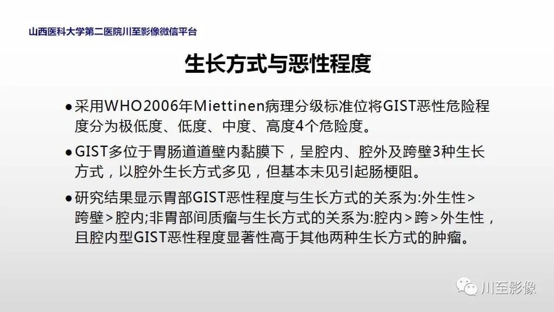
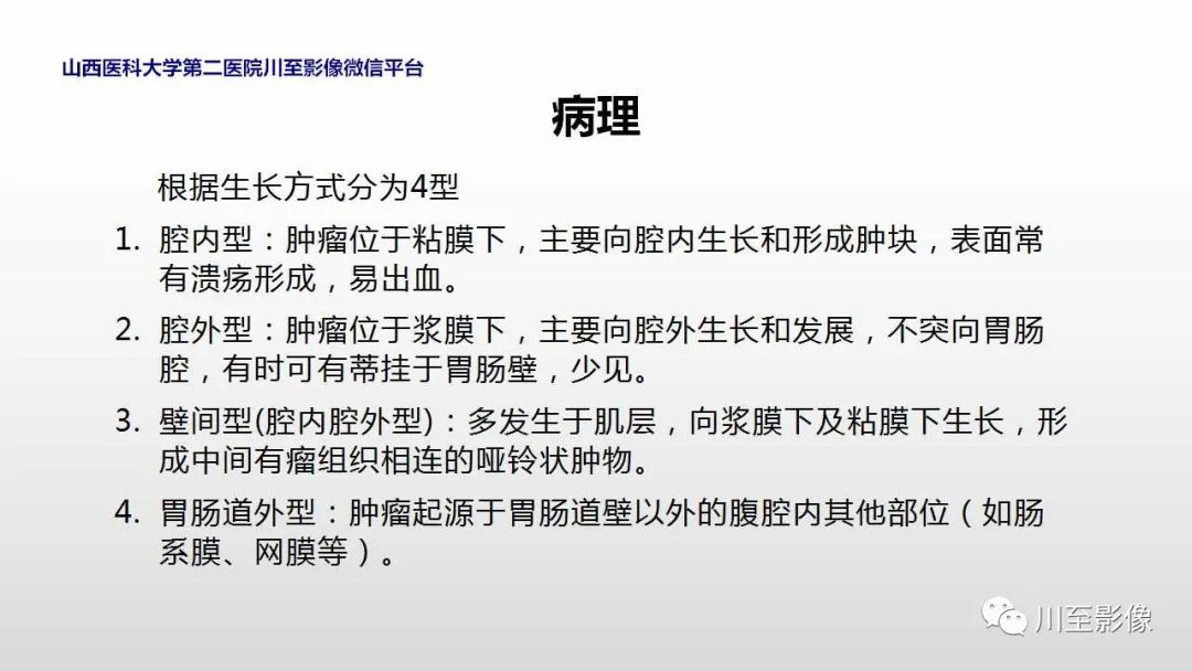
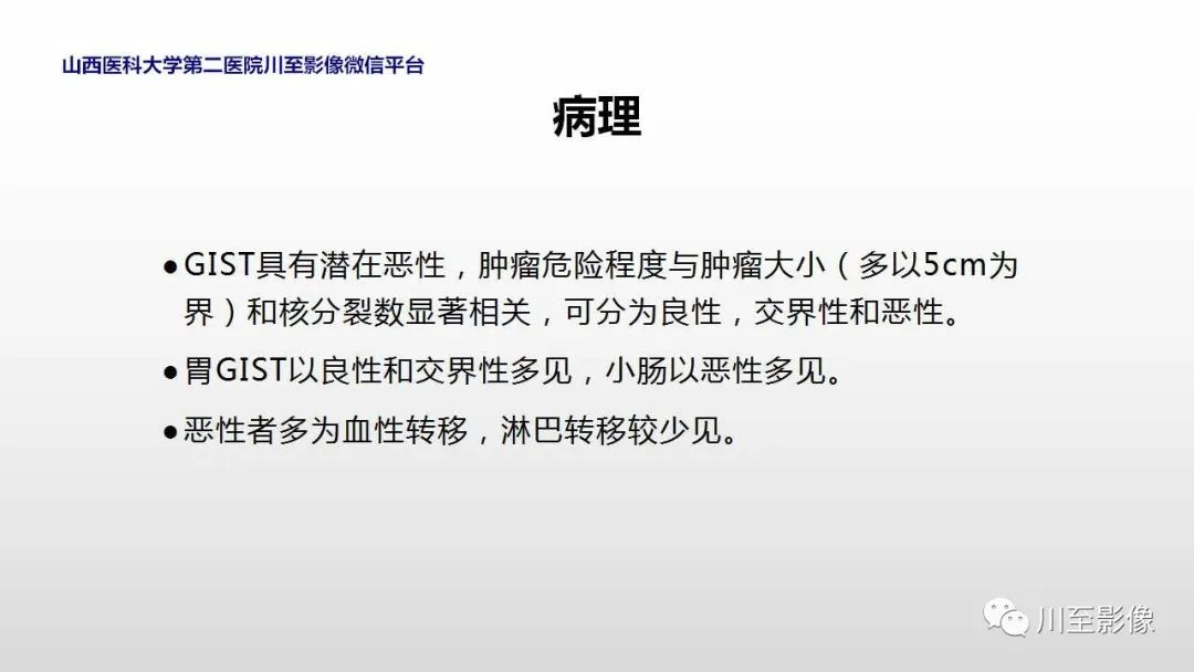
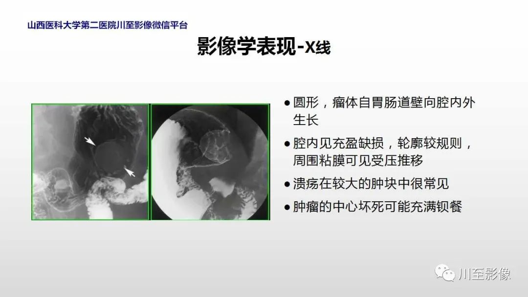
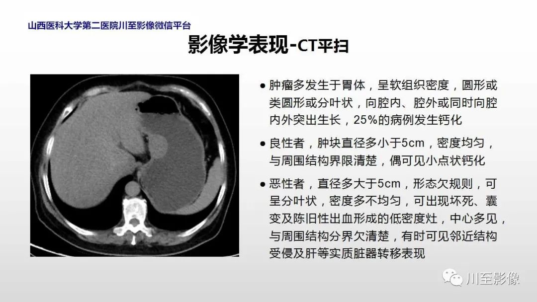
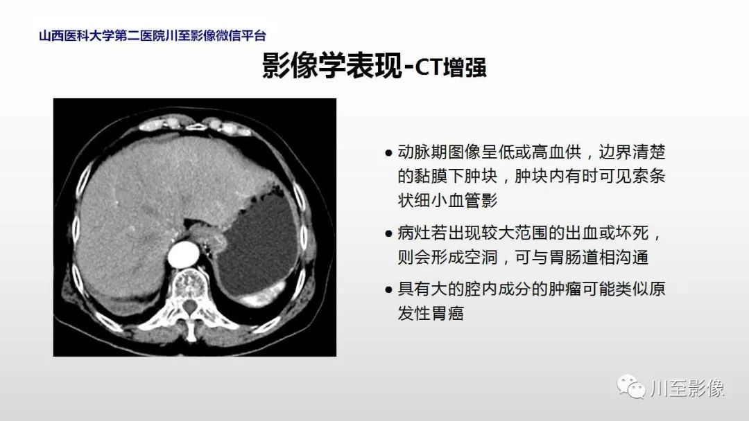
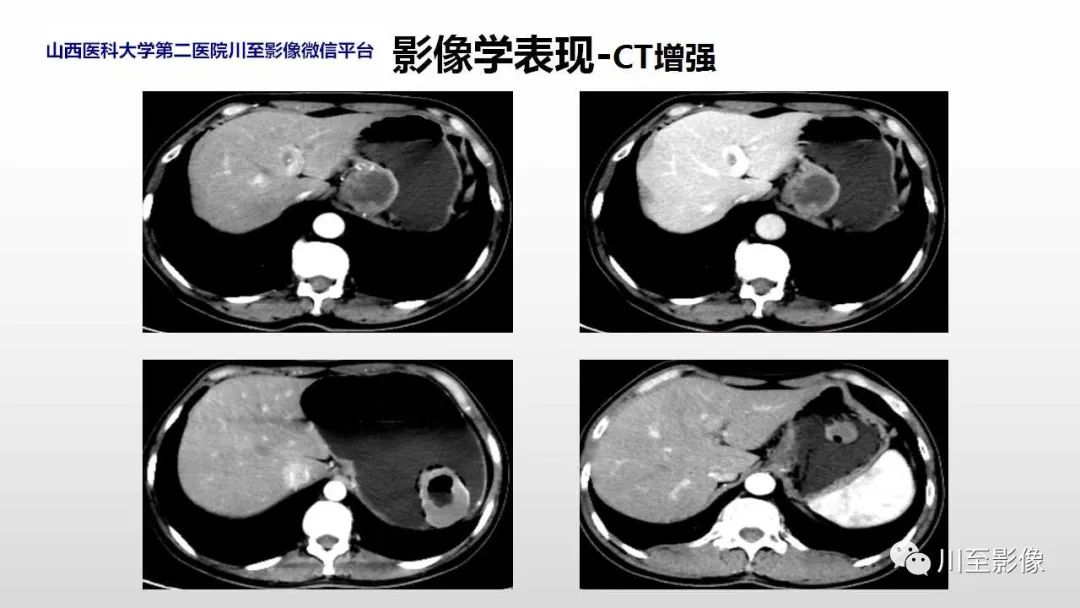
采用WHO2006年Miettinen病理分级标准位将GIST恶性危险程度分为极低度、低度、中度、高度4个危险度。GIST多位于胃肠道道壁内黏膜下,呈腔内、腔外及跨壁3种生长方式,以腔外生长方式多见,但基本未见引起肠梗阻。研究结果显示胃部GIST恶性程度与生长方式的关系为:外生性>跨壁>腔内;非胃部间质瘤与生长方式的关系为:腔内>跨>外生性,且腔内型GIST恶性程度显著性高于其他两种生长方式的肿瘤。
影像PPT - 【PPT】胃肠道间质瘤-25

发表回复

您的邮箱地址不会被公开。 必填项已用 * 标注