比萨综合征(Pisa syndrome)是一种罕见的临床病变,其特征在于躯干的明显的侧向弯曲,通常在运动时候表现明显,在仰卧位时缓解。在临床上,常见于潜在致残的神经退行性疾病,如帕金森病,多系统萎缩和阿尔茨海默病。比萨综合征发病机制尚不十分清楚,其治疗仍很棘手。
Pisa syndrome is a rare clinical entity characterized by marked lateral flexion of the trunk, which is typically mobile and resolves at supine position. When observed in clinical practice, it denotes an incapacitating symptom of underlying neurodegenerative disease such as Parkinson’s disease, multiple system atrophy, and Alzheimer’s disease. The underlying pathophysiological mechanisms responsible for the development of Pisa syndrome are poorly understood, and its management remains a challenge.
影像学表现:
站立位脊柱摄片,脊柱呈侧弯旋转畸形,仰卧位摄片或CT,大多数病人能缓解;CT可以显示肌肉萎缩伴脂肪变性。
推荐影像学评估:
- X-ray of the spine for calculating Cobb’s angle according to Cobb’s method;
- Computerized tomography (CT) scan of the dorsolumbar spinal muscles.
- Conventional radiographic investigation of the spine with the patients in the upright position in the anterior-posterior and lateral projections. CT scans of the lumbar portion of the spine were performed with patients lying in the supine position
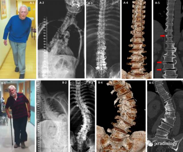
图A1-5站立位摄片及仰卧位CT重组,仰卧位病人侧弯能缓解;图B1-5,因为脊柱退变厉害,仰卧位侧弯不能缓解。具体请参考英文描述。—DOI: 10.1136/jnnp-2013-306573.152
Figure 1 Mobile and fi xed scoliosis in Pisa syndrome. Patient A had scoliosis on standing radiograph (A-2) but not when he was scanned supine (A-3, A-4 and A-5). There was evidence of osteophytic overgrowth below the apex of the scoliosis in the lumbar spine and above on the opposite side in the thoracic spine (A-4 and A-5), this pattern suggests the degenerative changes were working to stabilise his spine but stopped short at the apex of his curve leaving him mobile but tilted at that level when standing (A-1 and A-2). The reduction in curve with position, presence of interdiscal gas (red arrows A-5) and gaps between the osteophytes are evidence that despite attempts the deformity is not fi xed. Patient B had only minor improvement of his scoliosis on supine positioning (9% reducibility) (B-2 and B-3). Fusion of vertebral segments due to complete osteophytic bridging at the apex of the curve was clearly seen (B-4 and B-5) resulting in a fi xed and possibly stable spinal deformity.
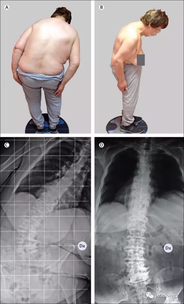
––Lancet Neurol 2016; 15: 1063–74
Figure 2: Clinical appearance of a patient with Parkinson’s disease and Pisa syndrome
(A and B) 69-year-old female patient with Parkinson’s disease and Pisa syndrome with forward fl exion of the trunk (mixed deformity), with a predominantly coronal plane deformity. (C) Anteroposterior radiograph of the same patient standing, showing right leaning of the trunk. Note that the spinous processes are aligned without rotation of vertebral bodies, thus diff erentiating Pisa syndrome from scoliosis. The lines provide a goniometric reference.(D) Supine projection showing a complete resolution of Pisa syndrome while lying down. Dx=patient’s right side.
来源:jxradiology