Histology
病理
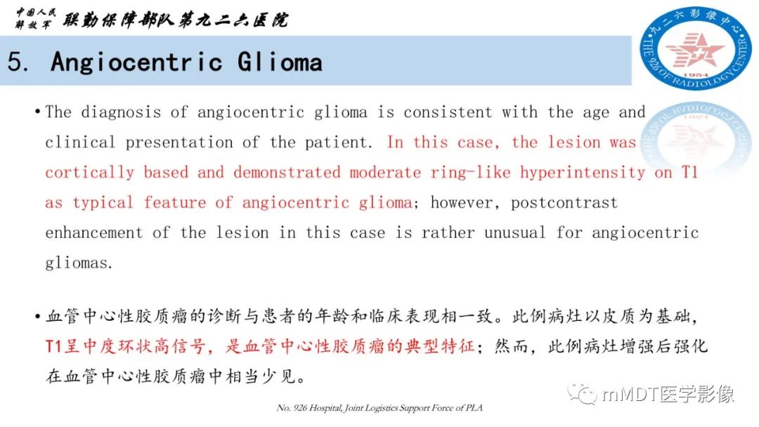
•The department of neuropathologyreceived a biopsy sample from a pediatric patient with therapy refractoryepilepsy. Histologically, the specimen appeared as a relatively isomorphicglial tumor of intermediate cell density (Fig. 5a). The tumor cell nuclei wereoval-shaped with intermediate chromatin density with a characteristic “salt andpepper” structure. No mitotic figures were found in 10 randomly selectedhigh-power fields. Nucleus-free perivascular zones were found throughout thetumor (Fig. 5a).
•神经病理科收到一名儿童难治性癫痫患者的活检样本。组织学上,标本表现为相对同构的中等细胞密度的胶质瘤(图5A)。肿瘤细胞核呈椭圆形,染色质密度中等,呈典型的“盐和胡椒”结构。随机选择的10个高倍视野均未发现有丝分裂影。肿瘤内可见无核血管周围带(图5A)。
Diagnosis–AngiocentricGlioma (WHO Grade I)
诊断-血管中心性胶质瘤(WHO I)
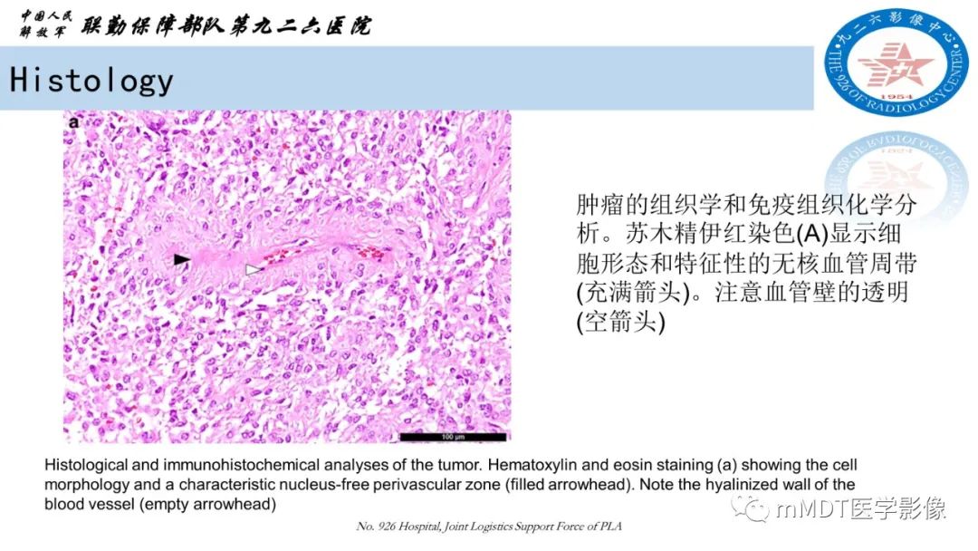
•Angiocentric gliomas areepilepsy-associated pediatric low-grade gliomas that display an angiocentricgrowth pattern with bipolar tumor cells and features associated with ependymaldifferentiation. This entity was first defined in 2005. Due toa morphological similarity to ependymomas, these tumors have previously beenreferred to as cortical ependymomas;however, a recent genome-wide methylation profiling analysis showed a cleardistinction between ependymomas and angiocentric gliomas.
•血管中心性胶质瘤是与癫痫相关的儿童低级别胶质瘤,呈血管中心性生长模式,肿瘤细胞两极,特征与室管膜分化相关。该实体首次定义于2005年。由于形态上与室管膜瘤相似,这些肿瘤以前被称为皮质室管膜瘤;然而,最近的全基因组甲基化图谱分析显示室管膜瘤和血管中心性胶质瘤有明显的区别。