Description
A 53-year-old man with history of recurrent headaches and recent onset of pulsatile tinnitus
53岁男性,既往有头痛病史。近期出现搏动性耳鸣。

Legends
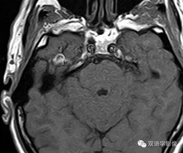
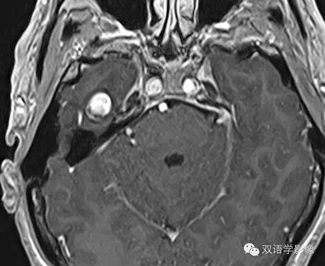
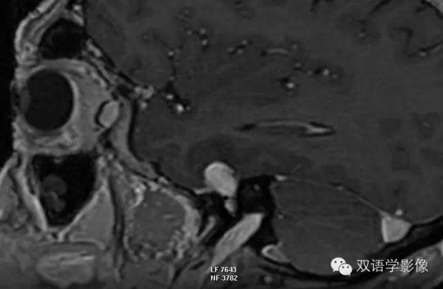
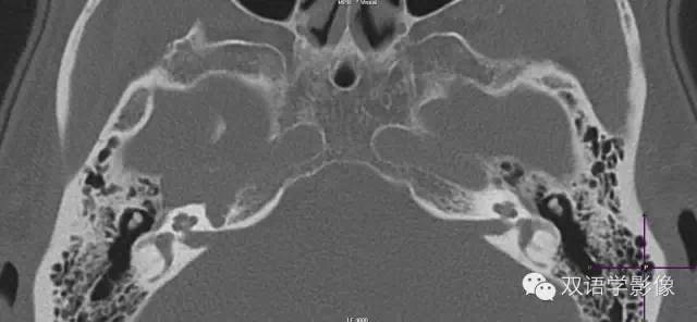
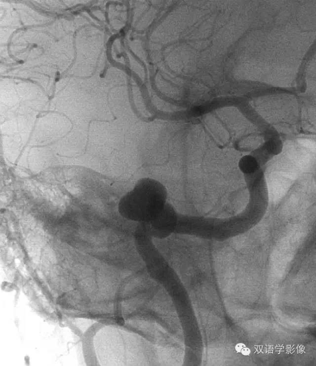
Coronal T2WI (A) demonstrates a well-defined ovoid hypointense lesion in the right temporal lobe. Axial T1WI (B) and corresponding fat-suppressed contrast-enhanced axial T1WI (C) demonstrate the lesion to be intrinsically hyperintense and showing near-homogenous enhancement. Sagittal T1WI (D) demonstrates the lesion to be clearly extra-axial. Noncontrast CT scan (E) demonstrates an expansile lesion in the region of the right petrous pyramid, which cannot be distinguished separately from the horizontal carotid canal. DSA (F) confirms a saccular aneurysm arising from the horizontal petrous segment of the right internal carotid artery.
冠状位T2WI示右侧颞部边界清楚的类圆形低信号。
横断位T1WI及对应的脂肪抑制T1WI增强扫描示病灶呈稍高信号,增强扫描均匀强化。
冠状位T1WI示示病灶位于脑外。
CT平扫示右侧颞骨岩部膨胀性病变,与颈动脉孔分界不清。
DSA证实为右侧颈内动脉岩段囊状动脉瘤。
Petrous Internal Carotid Artery Aneurysm
颈内动脉岩段动脉瘤
Petrous internal carotid artery (ICA) aneurysms are uncommon.
颈内动脉岩段动脉瘤临床较少见。
Clinical Presentation
Tinnitus, headache, and occasionally, hearing loss
耳鸣、疼痛,偶尔可伴有听力丧失。
Key Diagnostic Features
•MR imaging features demonstrate a ovoid enhancing lesion in the petrous pyramid.
MR表现为岩部类圆形结节,增强可强化。
•CT scan demonstrates an expansile lesion that cannot be distinguished separately from the horizontal petrous ICA.
CT可表现为膨胀性病变,与颈内动脉岩段分界不清。
•Peripheral calcification when associated with the lesion can help suggest the diagnosis.
如果合并钙化时,可有助于诊断。
•MR angiogram, CT angiogram, DSA can be diagnostic.
MRA、CTA、DSA可确诊本病。
DDx
•Cholesterol granuloma
胆固醇肉芽肿
•Schwannoma
许旺氏瘤
•Meningioma
脑膜瘤
•Paraganglioma
神经节细胞瘤
•Metastasis
转移瘤
•Lymphoma
淋巴瘤
•Leukemia
白血病
•