History: A 30-year-old woman presents with vertigo.
病史:30岁女性,眩晕。
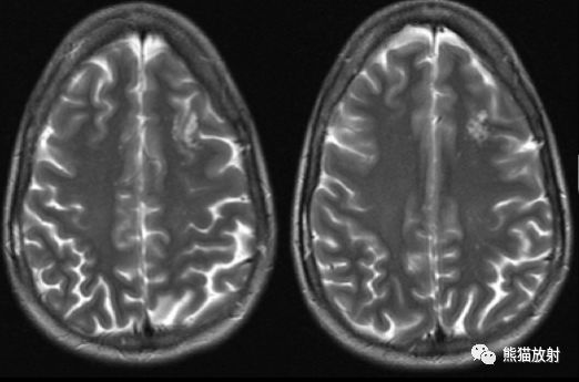
A contrast-enhanced MRI scan of the brain was performed. Axial T2-weighted, T2-weighted fluid-attenuated inversion-recovery (FLAIR), and T1-weighted and T1-weighted postcontrast MR images are shown below. 颅脑MR增强扫描:轴位T2WI、T2-FLAIR、T1WI及T1WI增强图像如下所示。


Findings
MRI demonstrates a cluster of well-delineated, variable-sized FLAIR/T2-hyperintense cysts within the left frontal subcortical white matter, without obvious mass effect or postcontrast enhancement.
影像表现:
MRI示左侧额叶皮层下白质内有一簇边界清晰的大小不等的FLAIR/T2高信号的囊肿,无明显占位效应或强化。
Differential diagnosis
- Multinodular and vacuolating neuronal tumor
- Dysembryoplastic neuroepithelial tumor
- Focal cortical dysplasia
- Tumefactive perivascular spaces
- Pilocytic astrocytoma
- Ganglioglioma
- Pleomorphic xanthoastrocytoma
鉴别诊断:
- 大脑多结节及空泡性神经元肿瘤
- 胚胎发育不良性神经上皮肿瘤(DNET)
- 局灶性皮质发育不良
- 瘤样血管周围间隙
- 毛细胞型星形细胞瘤
- 节细胞胶质瘤
- 多形性黄色星形细胞瘤
Diagnosis: Multinodular and vacuolating neuronal tumor (MVNT)
最后诊断:大脑多结节及空泡性神经元肿瘤 (MVNT)
MVNT
Pathophysiology, epidemiology, and clinical presentation:
Multinodular and vacuolating neuronal tumors are a more recently characterized World Health Organization (WHO) grade 1 central nervous system neoplasm. This rare entity has only been described in several case series. They are characterized as a benign, mixed glial neuronal lesion consisting of nodules of disorganized neuronal cells located near the subcortical white matter. They are associated with long-term seizures in adults in about 30% of patients, but they are most often found incidentally and are considered “do not touch” or “leave me alone” lesions if not associated with intractable epilepsy.
病理生理学,流行病学和临床表现:
多结节和空泡性神经元肿瘤是最近出现在中枢神经系统肿瘤WHO1级中的,这种罕见的病变仅在几个案例系列中被描述。
主要特征是良性的混合性的胶质神经元病变,位于皮层下白质附近,由紊乱的神经元细胞结节组成。
与大约30%的成年人的长期癫痫发作有关,通常为偶然发现,如果与难治性癫痫无关,无需处理。

Imaging features
- They are difficult to identify on CT but may exhibit hypoattenuation.
- On MRI, they are seen as a subcortical cluster of tiny, cystic, nodular lesions with associated T2/FLAIR signal abnormality (and mildly T1 hypointensity).
- They are usually seen in the deep cortical ribbon and superficial subcortical white matter.
- They rarely enhance (approximately 3% to 10% of cases) with stippled faint enhancement.
- They should not exhibit restricted diffusion or abnormal susceptibility.
- They rarely progress/increase in size.
影像表现:
- 在CT上很难明确病变,可表现为低密度;
- 在MRI上表现为T2 / FLAIR信号异常(T1轻度低信号)的皮层下簇状微小囊性结节性病变;
- 多见于深部皮质带和浅表皮层下白质;
- 多无强化,约3%-10%的病例可见点状微弱增强;
- 无扩散受限或异常磁敏感性;
- 病灶大小几乎不会进展/增加。
Treatment
They are slow-growing/indolent, and if not associated with refractory epilepsy, they are considered a “leave me alone” lesion.
They are usually just followed with imaging, but they can be resected if they are associated with intractable epilepsy.
治疗:
- 病灶生长缓慢或无变化,如果不与顽固性癫痫相关,被认为是“别管我”病灶。
- 通常借助影像进行随访,如果与难治性癫痫相关则予以切除。