Fig 1.11.1

Fig 1.11.2
HISTORY:
21-year-old woman with aliver mass identified incidentally on CT during workupfor acute appendicitis
21岁女性,因阑尾炎行CT检查,发现肝脏肿块。
IMAGING FINDINGS:
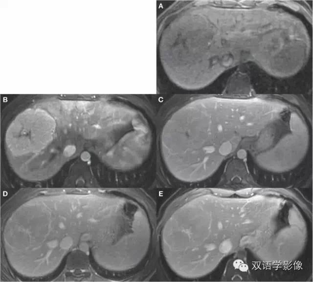
Axial fat-suppressed FSE T2-weighted image (Figure 1.11.1)demonstrates a large right hepatic lobe mass with mild hyperintensity relative to liver and a high intensity central scar. IP and OP T1-weighted 2D SPGR images showed no signal dropout to suggestintravoxel fat (images not shown). Pregadolinium and arterial, portalvenous, equilibrium, and 5-minute delayed phase postgadolinium 3D SPGR images (Figure1.11.2) demonstrate marked,uniform arterial phase enhancement within the lesion that rapidly becomes isointensewith liver. Note also gradual enhancement of the central scar, as well as peripheral rim enhancement.
横断位T2WI FSE脂肪抑制序列(Figure 1.11.1)示肝右叶巨大稍高信号肿块肿块,内可见中央瘢痕,呈明显高信号。2D SPGR T1WI同反相位图像未见明显信号衰减,提示病灶内没有脂肪成分(图像未示出)。3D SPGR增强扫描动脉期、门静脉期、平衡期及5分钟延迟扫描(Figure1.11.2)示动脉期明显均匀强化,然后信号迅速降低,门静脉期及平衡期病灶与肝实质呈等信号。另外,中央瘢痕和病灶边缘呈渐进性强化。
DIAGNOSIS:
Focal nodular hyperplasia
局灶性结节增生
COMMENT:
FNH is the second most common benign tumor of the liver after
hemangiomas, accounting for 8% of these lesions and with an estimated prevalence of 0.9%。FNHs consist of hyperplastic hepatocytes and small bile ductules surrounding a fibrovascular central scar that is thought to represent a hyperplastic response to a preexisting vascular malformation rather than a true neoplasm. They are more prevalent in women(usually of reproductive age) than men by an 8:1 ratio and are solitary in 80% to 95% of cases. Since FNHs are benign non-surgical lesions, the most important job of the imager is to make a confident diagnosisand distinguish them from more potentially unfriendly hypervascular lesions, such as adenoma, HCC, and metastases. Most of the time, this is relatively easy to accomplish.
FNH是仅次于血管瘤的肝脏第二常见良性肿瘤,约占全部肝肿瘤的8%,发病率约为0.9%。FNH主要由增生的肝细胞、小胆管包绕中心纤维血管瘢痕构成,一般认识是血管畸形基础上的增生性病变,而非真正的肿瘤。FNH女性多于男性,男女比例约8:1,且通常发生于育龄妇女。单发病灶多见,约占全部的80%-95%。由于FNH是良性病变,不需要手术治疗,所以对于放射科医师来说,最主要的任务是明确诊断,与其他富血供潜在恶性肿瘤相鉴别,如腺瘤、HCC、转移瘤等。通常FNH的诊断和鉴别诊断并不难。
Classic FNHs (illustrated by this case) are described as invisible or barely perceptible lesions except on postgadolinium arterial phase images; they are typically isointense to liver on T1-weighted images and isointense or mildly hyperintense on T2-weighted images. It is true, however, that FNHs are often at least moderately hyperintense on diffusion-weighted images, which illustrates the general principle that DWI is a technique better used for lesion detection than lesion characterization.Dynamic postgadolinium images show intense uniform enhancementon arterial phase images, which quickly becomes nearly isointense to liver on portal venous and equilibrium phaseimages. The central scar (seen in this case but not universally identifiable) generally shows high signal intensity on T2-weighted images and gradual enhancement following gadolinium administration. An elaborate set of imaging characteristics of the central scar has been described in the literature as a means of distinguishing FNH from fibrolamellar hepatoma; however, the percentage of lesions thatdon’t follow the rules is high enough that we rarely find these guidelines particularly useful.
典型的FNH(如本例患者)在T1WI呈等信号,T2WI呈等或稍高信号,所以除了增强扫描动脉期外,有时难以发现。然而在DWI图像上,病灶通常呈高信号,说明DWI在病灶的发现上作用大于定性诊断。动态增强扫描,动脉期病灶明显强化,门脉期及延迟期病灶与肝实质呈等信号。病灶中央瘢痕(本例患者可见,但并非全部可见)常呈T2WI高信号,增强扫描渐进性强化。中央瘢痕的这种典型表现通常作为FNH与纤维板层样肝癌的鉴别点,但很多的病例的表现并不如此典型,所以这一原则并非特别实用。
This case illustrates the appearance of a very large, but otherwise typical, FNH,obtained with a standard extracellular gadolinium-based contrast agent. The examination was done several years ago and exemplifies one of the problems of advancing technology: We read this case as consistent with FNH and offered no differential diagnosis. If it were to appear today,however, the report likely would containa differential that included adenoma (the hedge isthe radiologist’sfavorite plant) and suggest that a follow-up examination with a hepatobiliarycontrast agent be performed (although the absolutely classic appearance of the central scar might provide enough assurance for bold radiologists to remain firm in their conviction). This is entirely unnecessary; we used to be very certain about what FNH looked like usingstandard extracellular gadolinium contrast agents and were rarely mistaken. Although it is true that small adenomas can have a nearly identical appearance to FNH, they aren’t verycommon and it’s not clear whether missing one of these lesions results in any actual harm.
此例患者病灶体积较大,但细胞外造影剂增强扫描图像非常典型。此例患者是几年前采集的,也说明了影像技术进步的一个问题:当时我们看到这个病例,考虑为FNH,并没有提供鉴别诊断。而如果现在看到这个病例,诊断报告中可能会提出鉴别诊断,包括腺瘤,并且建议肝胆特异性对比剂增强进一步检查(尽管典型的中心瘢痕的表现足以让胆大的放射科医师坚持做出诊断)。这完全没有必要。我们已经非常了解FNH在细胞外对比剂增强扫描中的表现,很少会诊断错误了。尽管较小的腺瘤可以表现非常类似于FNH,而且没有证据表明如果混淆这两者,会有什么实际性的伤害。
This case demonstrates another well-established principle: Clinicians (and patients)don’tlike large masses, regardless of whatyou say about them. Despite a confident radiologicdiagnosis, as well as an absence ofsymptoms or abnormal laboratory values, much discussion ensued regarding the possibility of fibrolamellar hepatoma and the virtues of resection. Fortunately, surgery was avoided, but an 8-month follow-up examination was performed, which had exactly the same appearance as the first one.
这个病例也说明了另外一个原则:无论你对他们说什么,临床医师(和患者)不喜欢大肿瘤。尽管放射科医师对诊断非常有信心,且患者没有任何症状,实验室检查也基本正常,但还是对纤维板层样肝癌的可能性及手术的优点进行了很多次讨论。幸运的是,该患者并没有进行手术,但还是进行了长达8个月的随访,表现与第一次完全相同。
Thecase was also one of the first abdominal MRI examinations performed at MayoClinicwith a 3-T magnet.A 3-T system has many real and some theoretical virtues. SNR is proportional to magnetic field strengthand therefore in theory is doubled at 3 T (in practiceit’s usually a little less than this), and the improved signal strength can be used as currency to purchase higher spatial resolution with preserved SNR or faster scans (ie, fewer signalaverages or higher acceleration factorswith parallel imaging).
这里患者也是Mayo Clinic第一例3.0T腹部MRI。3.0T的系统从理论上和实际上有着很多优点。从理论上来说,SNR与场强成正比,所以3.0T磁共振SNR是原来的2倍(实际上比这个略小一点),信号强度的增高可以用来保证信噪比及缩短扫描时间(如减少平均采集次数或使用并行采集技术)的同时,获得更高的空间分辨率。
There are many limitations, however, including longer T1 and slightly shorter T2 relaxation times, meaning moresaturation (and lower signal) for the same TR and more rapidsignal decay (and lower signal) for thesame TE. More importantly, susceptibility artifactsare greatly increased, as are standingwave and conductivity artifacts, which can lead to severe signal loss in all or part ofthe image. Although 3-T MRI has become a part of our daily practice and has generated somespectacular images, it is also true that we occasionally need to transfer patients back to a1.5-T magnet because the 3-T images are uninterpretable。
3T的MRI同样也有一定的不足。更长T1、更短T2在相同TR时意味着更加饱和(更低信号);使用相同TE,也有更多的信号衰减(更低信号)。更重要的是,3.0T磁共振的磁敏感伪影更加明显,“驻波效应”和“传导性效应”也会增加,会导致全部或部分图像信号损失。尽管3.0T的磁共振已经逐渐应用于临床,且能够获得更加清晰的图像,但有时由于图像无法接受,我们不得不让病人重新进行1.5T磁共振检查。