History: A 75-year-old man presents with headaches.
病史:老年男性,75岁,头痛。
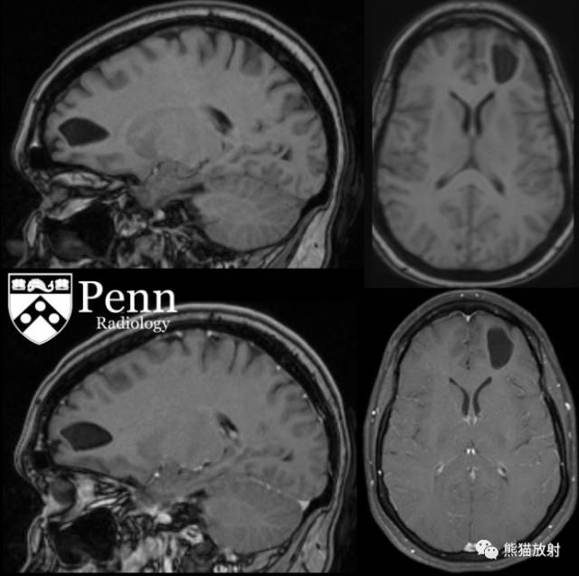
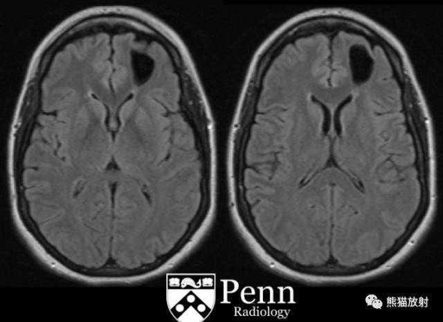
Sagittal and axial T1 pre- and postcontrast, axial T2-weighted, and axial fluid-attenuated inversion-recovery (FLAIR) MR images are shown below.
矢状、轴位T1平扫及增强,轴位T2WI、轴位FLAIR图像如下所示。

Findings
There is a simple, intra-axial cyst with smooth contours in the anterior left frontal white matter measuring up to 2.9 cm. There is no abnormal enhancement or surrounding parenchymal signal abnormality. There is no communication to the ventricle or superficial subarachnoid space.
影像表现:
左额叶白质见一单纯脑内囊肿,轮廓光滑,大小约2.9cm。增强无强化,无周围水肿。不与脑室或表面的蛛网膜下腔相交通。
Differential diagnosis
Neuroglial cyst
Dilated perivascular space
Subarachnoid cyst
Porencephalic cyst
Neurocysticercosis
Cystic neoplasm/low-grade gliomas
- Dysembryoplastic neuroepithelial tumor
- Pilocytic astrocytoma
- Pleomorphic xanthoastrocytoma
- Multinodular and vacuolating neuronal tumor
Cystic periventricular leukomalacia
鉴别诊断:
神经上皮囊肿
扩大的血管周围腔隙
蛛网膜囊肿
脑穿通囊肿
脑囊虫病
囊性肿瘤/低级别胶质瘤
- 胚胎发育不良性神经上皮肿瘤
- 毛细胞型星形细胞瘤
- 多形性黄色星形细胞瘤
- 多结节和空泡神经肿瘤
囊性脑室周围白质软化症
Diagnosis: Neuroglial cyst
最后诊断:神经上皮囊肿
Discussion
Neuroglial cyst
Pathophysiology
Neuroglial cysts, also called glioependymal cysts, are benign epithelial-lined lesions that can be found anywhere along the neuroaxis. They can be extraparenchymal or intraparenchymal, that latter of which are more common. Intraparenchymal cysts are congenital lesions arising from embryonic neural tube elements sequestered in the developing white matter. They can be lined by either ependymal epithelium (columnar epithelium) or choroid plexus cells (low cuboidal epithelium). They do not communicate with the ventricular system or arachnoid space.
病理生理学
神经上皮囊肿,也称为胶质室管膜囊肿,是良性内衬上皮病变,可见于神经轴索走行的任何部位。
可发生在脑外或脑内,后者更多见。
脑内囊肿是先天性病变,其起源于白质发育过程中退化的胚胎神经管,可内衬室管膜上皮(柱状上皮)或脉络丛细胞(立方上皮)。
囊肿不与脑室或蛛网膜下腔相通。
Epidemiology
They are uncommon, representing only 1% of intracranial cysts. They are usually found incidentally and do not cause any symptoms.
流行病学
少见,仅占脑内囊肿的1%,通常为偶然发现,不导致任何症状。
Imaging features
- Neuroglial cysts appear as round, smooth, unilocular cysts containing clear fluid.
- They can be seen anywhere, with the most common location being the frontal lobes.
- They are usually solitary (in contrast to dilated perivascular spaces).
- They follow cerebrospinal fluid signal characteristics on all sequences.
- They do not show abnormal enhancement.
- They do not show restriction of diffusion.
影像表现
- 神经上皮囊肿表现为类圆形,边缘光滑,单房囊肿,内包含清亮液体;
- 可见于脑内任何部位,最常见于额叶;
- 通常为单发(可与扩大的血管周围腔隙鉴别);
- 病变在MR所有序列上均呈脑脊液信号;
- 无异常强化;
- 无弥散受限。