Epidermoed 表皮样囊肿
Arachnoid Cyst 蛛网膜囊肿
Cholesterol Granuloma 胆固醇肉芽肿
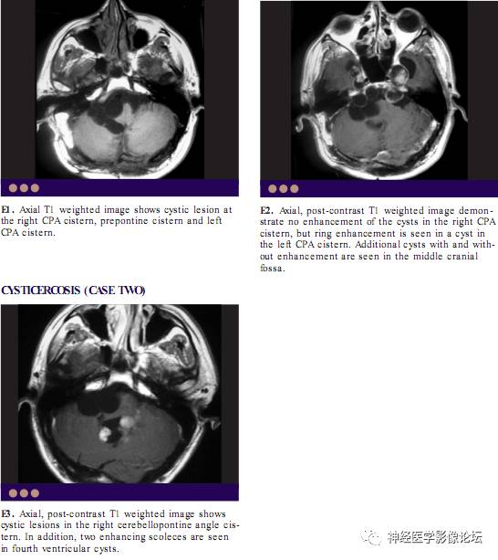
Cysticercosis Cyst 猪囊尾蚴囊肿
Epidermoed
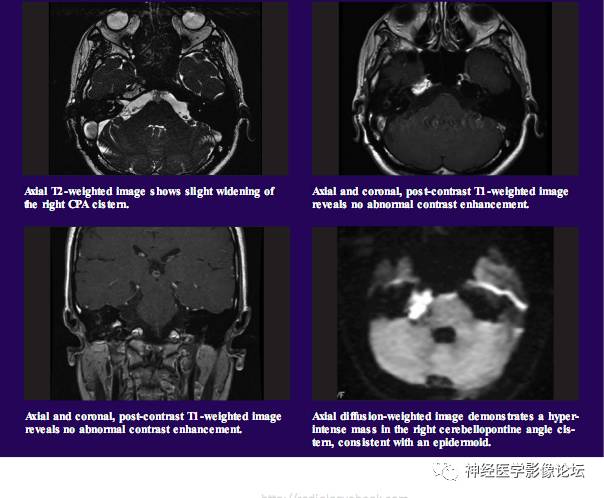
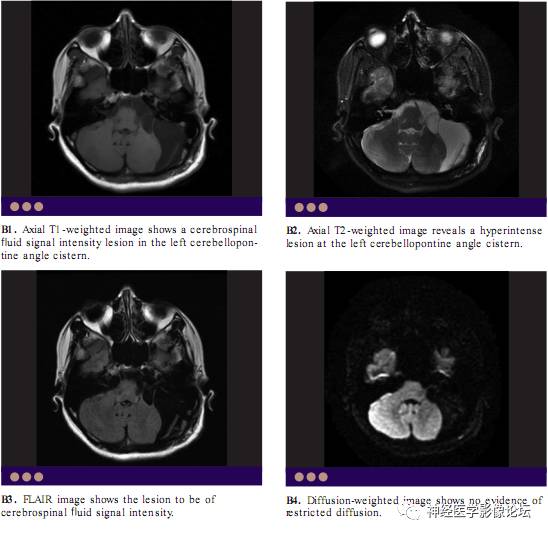
Epidermoids are congenital masses, but their clinical presentation usually in the third or fourth decade. They are predominantly located in basal cisterns and are usually paramedian in location as opposed to the midline location of dermoids. Epidermoids are the third most common masses of CPA. Epidermoids have similar signal intensity features to those of CSF on both T1-weighted and T2-weighted images. Sometimes, an internal stranded appearance can be seen, giving a “dirty-CSF”appearance. FLAIR imaging may show a slightly hyperintense lesion as compared to CSF. Diffusionweighted images can be useful in differentiating epidermoids from arachnoid cysts. Epidermoids show restricted diffusion whereas arachnoid cysts do not show restricted diffusion.
Arachnoid Cyst
Cholesterol Granuloma
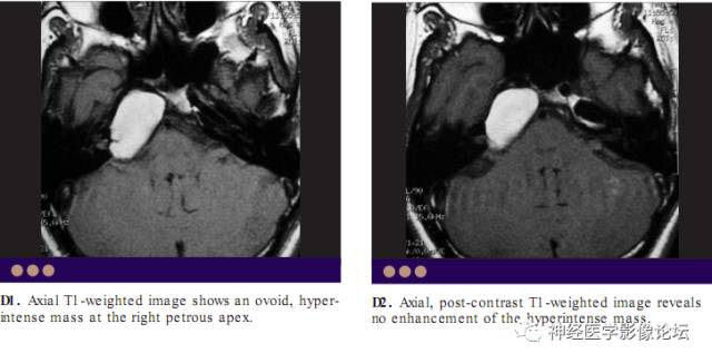
Cholesterol granuloma usually involves the petrous apex. Recurrent hemorrhage into the obstructed air cell at petrous apex with secondary inflammatory changes is thought to be the cause for cholesterol granuloma. Products of blood degradation, cholesterol crystals form a slowly expansile, fluid filled lesion at the petrous apex.
Cysticercosis Cyst
Cysticercosis cysts in the basal cisterns usually take the racemose form and they do not contain the scolex. Since they are cysts of CSF signal intensity, they are difficult to differentiate from arachnoid cysts or epidermoids. Sometimes,contrast enhancement may be seen adjacent to the cysticercosis cysts due to the presence of inflammatory reaction adjacent to these cysts. They are similar to arachnoid cysts and do not exhibit restricted diffusion.