History: A 46-year-old man presents to the emergency department (ED) with neck fullness and abdominal pain. The patient moved to the U.S. from Liberia 10 years ago.
病史:46岁男性,因颈部肿胀、腹部疼痛就诊于急诊科,患者十年前曾从美国去过利比里亚。
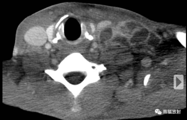
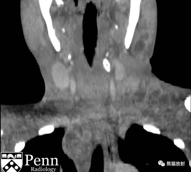
A contrast-enhanced CT scan of the neck was performed. Axial and coronal images are shown below.
行颈部CT增强,轴位及冠状位如下所示。
Additional images
Further imaging was obtained due to the patient\’s nonspecific abdominal discomfort.
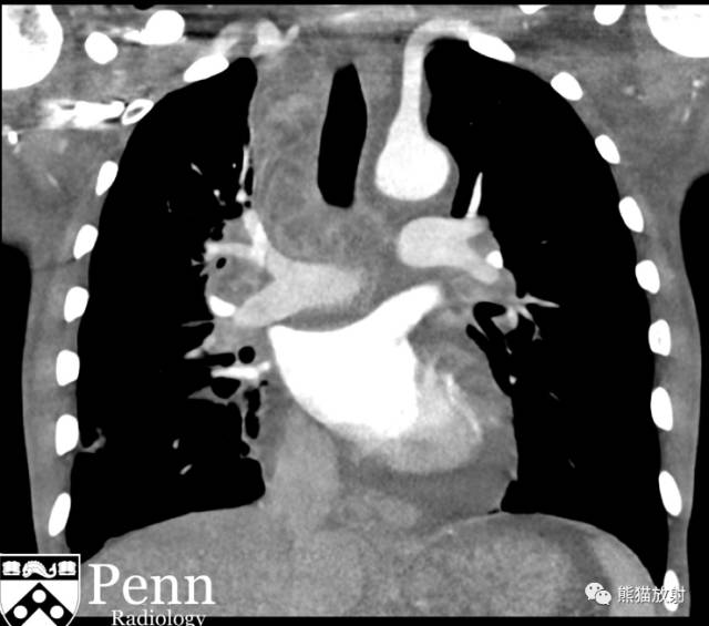
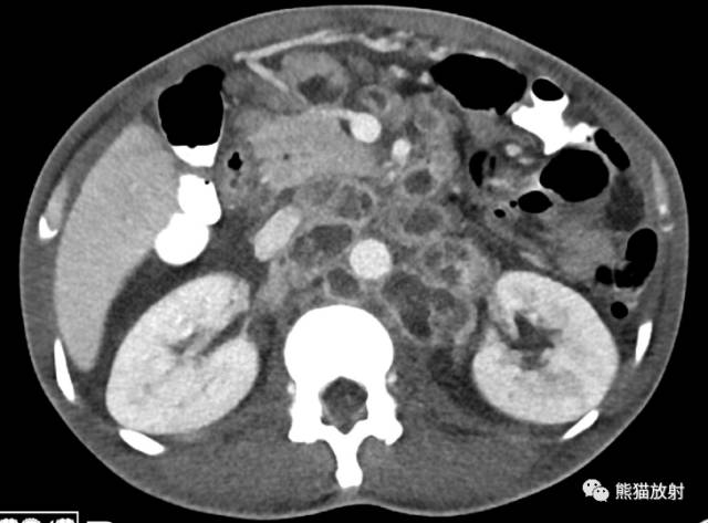
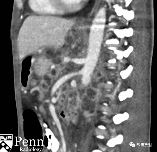
Contrast-enhanced CT scans of the chest and abdomen/pelvis were performed. Coronal, axial, and sagittal images are shown below.
由于患者腹部不适,进一步行胸腹盆CT增强,冠状、轴位及矢状图像如下所示。
Findings
Necrotic lymphadenopathy is seen through the cervical region, mediastinum, hila, abdomen, and retroperitoneum.
影像表现:
颈部、纵隔、肺门、腹腔及腹膜后见多发坏死性肿大淋巴结。
(补充:增强扫描呈环形强化)
Differential diagnosis
- Tuberculous lymphadenopathy
- Lymphoma
- Metastatic carcinoma
- Mycobacterium tuberculosis infection
- Nontuberculous mycobacterial infection
- Disseminated histoplasmosis infection
- Whipple disease
- Autoimmune disease (i.e., systemic lupus erythematosus)
鉴别诊断:
- 淋巴结结核
- 淋巴瘤
- 转移癌
- 结核分枝杆菌感染
- 非结核分枝杆菌感染
- 播散型组织胞浆菌病感染
- 惠普尔病
- 自身免疫性疾病(如:系统性红斑狼疮)
Diagnosis: Tuberculous lymphadenopathy, biopsy-proven
诊断:淋巴结结核(活检证实)
Key points
Tuberculous lymphadenopathy
Background
- Tuberculosis (TB) is typically confined to the respiratory system.
- However, it can affect any organ system, and immunocompromised patients have an increased risk of extrapulmonary TB.
- Radiographic evidence of lymphadenopathy is seen in more than 95% of children and over 40% of adults with TB.
- Lymphadenopathy as the only evidence of TB radiographically is more common in infants and decreases in frequency with age.
背景:
- 结核(TB)通常局限于呼吸系统;
- 然而,也可以累及其它器官,免疫缺陷人群更易患肺外结核;
- 超过95%的儿童及超过40%的成人结核患者影像学可发现淋巴结肿大;
- 婴儿常常仅表现为淋巴结肿大,这种情况随年龄增加而减少。
Imaging
CT:
- Lymph nodes larger than 2 cm often have a low attenuation center from necrosis and are suggestive of active disease.
- Cervical TB lymphadenopathy is often the first site of extrathoracic lymph node involvement; known as scrofula or cervical TB lymphadenitis.
影像表现(CT):
- 淋巴结大于2cm时常由于坏死而表现为中心低密度,提示疾病处于活动期;
- 颈部结核性淋巴结肿大常是胸外淋巴结受累的第一站,被称作淋巴结结核或颈部结核性淋巴结炎。
Diagnosis
- Outside of the U.S., up to 50% of patients with chronic lymphadenopathy have TB as the causative etiology. However, malignancy is a significant alternative diagnosis.
- Thus, a biopsy is thus recommended to confirm TB and exclude other diagnoses.
诊断:
- 在美国以外的地方,多达50%的慢性淋巴结肿大患者的病因为结核;然而,恶性肿瘤是一个重要的替代诊断;
- 因此,需活检证实为TB,除外其他诊断。
几例淋巴结结核、肿瘤
女,2岁,30余天前无明显诱因下出现左侧颈部肿物,起初小,约1×1cm,进行性增大,皮肤表面无红肿、发热,无溃烂,当地医师给予草药局部外敷,无明显疗效。
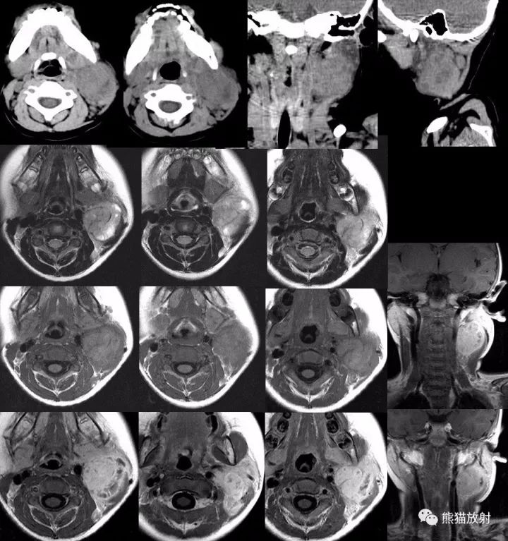
左颈部明显肿大淋巴结,密度、信号不均匀,内见斑点状无强化坏死囊变区,增强扫描大部分区域呈明显强化。
术后病理:颈部包块:灰白灰红色类圆形肿物一枚,大小4x3x3.3cm,切面灰黄灰红相间;颈淋巴结2枚,1枚米粒大,另一枚灰白灰红色,类圆形,大小1×0.6×0.3cm。送检颈部包块及颈部淋巴结见淋巴结结构,淋巴结组织内多处可见多个片状的干酪样坏死组织,周围可见较多由类上皮样细胞及郎罕氏巨细胞构成的结核结节,其间散在多少不等的淋巴细胞浸润。在病变组织边缘见少量横纹肌及脂肪组织,未见确切腮腺组织。
(左颈部)淋巴结结核
女,17岁,发现右侧颈部肿物十余天。
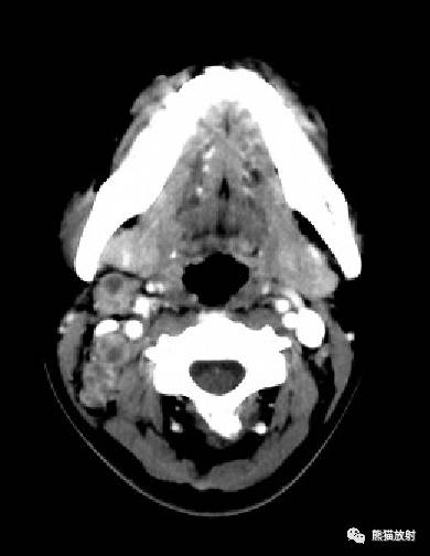
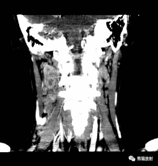
右颈部多发增大淋巴结,增强扫描均可见坏死区。
入院后完善检查,PPD试验( ),血沉明显加快,颈部包块细胞学活检示符合颈部淋巴结结核。
男,28岁,无意间发现右颈部包块,无疼痛,无发热、盗汗。
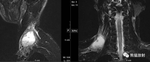
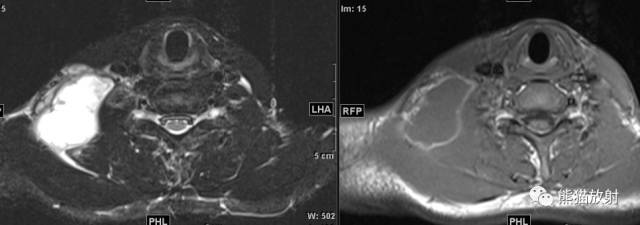
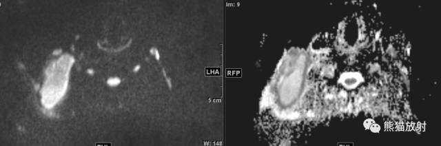
行手术切除,术后病理:淋巴结结核
男性,71岁,发现颈部包块1天。
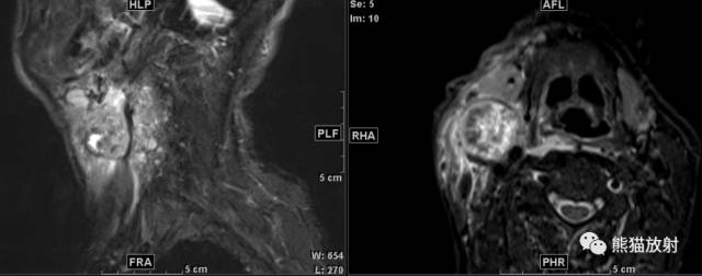
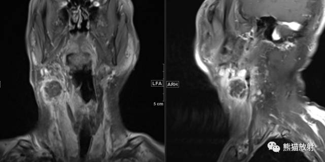
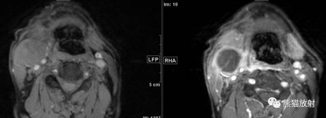
(颈部淋巴结穿刺)转移性鳞状细胞癌
男33岁,颈部无痛性肿物3月余,质硬,活动度差,轻度触痛。
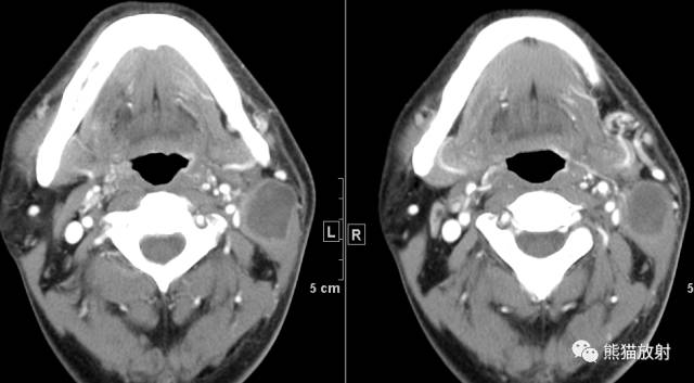
左颈部淋巴结鳞状细胞癌
鉴别诊断要点
- 年龄:结核多见于幼儿、青少年;转移癌多见于中老年;
- 全身情况:临床症状,有无肺结核(不一定有);有无原发肿瘤(不一定能找到);
- 影像表现:结核的坏死区相对边界较清楚,淋巴结之间边界相对清晰,无明显融合趋势;转移癌的坏死区多欠光整,淋巴结周围毛糙,易融合成团;
- 建议:病理学检查。