MENINGITIS
(脑膜炎)
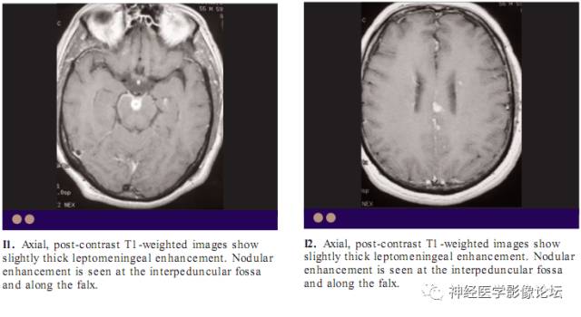
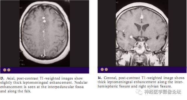
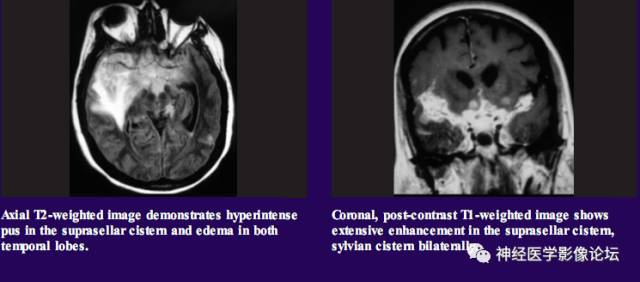
The primary mechanism of leptomeningeal enhancement in meningitis is breakdown of the blood–brain barrier(血脑屏障) without angiogenesis. Bacterial(细菌性) and viral(病毒性) meningitis typically show thin and linear enhancement pattern. Fungal(真菌) meningitis may produce thicker, lumpy, or nodular enhancement in the subarachnoid space .Aspergillus fumigatus infection is seen predominantly in immunocompromised(免疫功能低下) patients. CNS aspergillosis is more commonly caused by hematogenous spread (血行播散)of pulmonary disease and less commonly caused by direct extension of disease in the nasal cavity and paranasal sinuses. Pathologically, diffuse vascular invasion with thrombosis of cerebral vessels occurs . Necrotizing arteritis of small vascular channels is also seen.
CONTUSION
(挫伤)
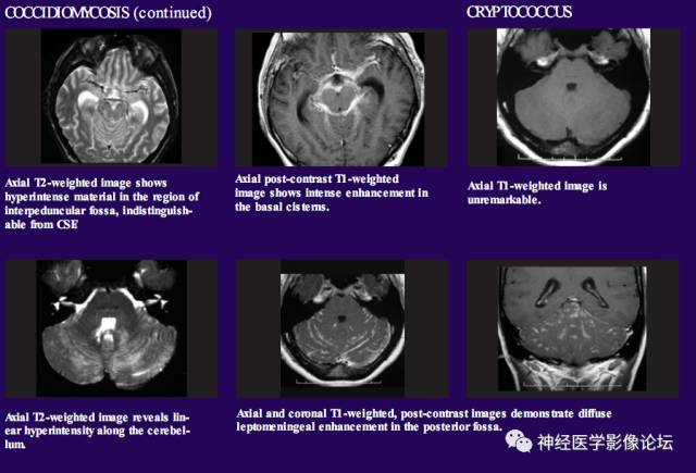
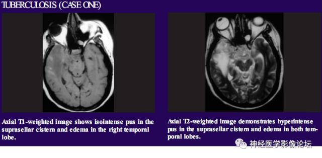
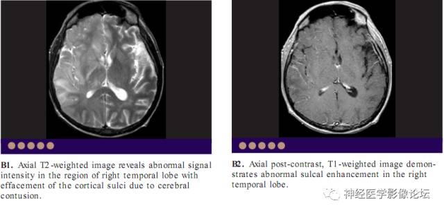
A contusion is a type of traumatic brain injury that causes bruising of the brain tissue. A contusion may result from a blunt trauma on the head that causes damage to the brain.The most common cause of traumatic brain injury(外伤性脑损伤) is motor vehicle accidents . Sports or physical activity is the second most common cause, and assaults are third. Leptomeningeal enhancement results from breakdown of the blood–brain barrier.
CYSTICERCOSIS
(囊虫病)
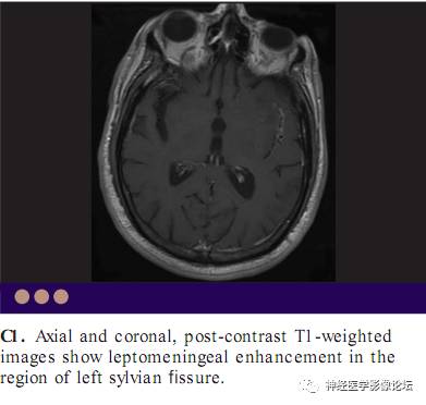
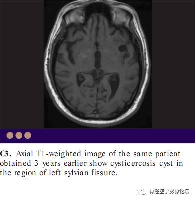
Intracranial cysticercosis has four types , namely, intraparenchymal, cisternal, ventricular, and mixed. When cisternal cysts degenerate in the subarachnoid space(蛛网膜下腔), they can cause leptomeningeal enhancement.
INFARCT
(梗塞)
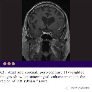
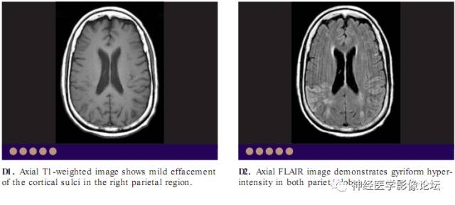
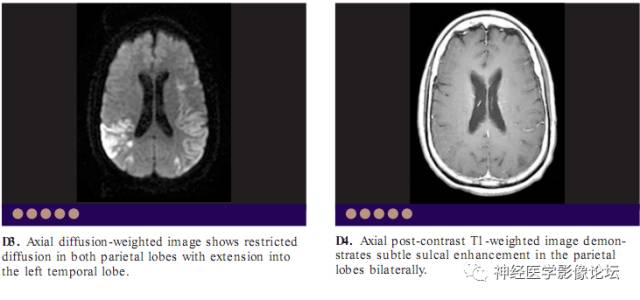
Detection of very early infarcts is better with MR imaging than with CT because of its superior contrast resolution. MR can detect is chemic infarcts in 96% of the cases with conventional MR scanning techniques in the first 24 hours. MR imaging may demonstrate the absence of flow void(流空) in a major vessel, loss of gray–white differentiation, focal edema, and leptomeningeal enhancement. MR diffusion imaging and MR perfusion imaging(灌注成像) are now regarded as the imaging techniques of choice because they permit accurate, reliable diagnosis and characterization of ischemic strokes within the critical first 6-hour time period needed to initiate thrombolytic therapy(溶栓治疗).
LEPTOMENINGEAL METASTASIS
(软脑膜转移)
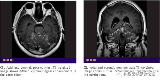
The enhancement due to leptomeningeal carcinomatosis can appear as multiple nodules, diffuse leptomeningeal enhancement, ependymal(室管膜) or subependymal(室管膜下) enhancement, dural enhancement, or a combination of the above. In the nodular form, pial enhancement is difficult to distinguish from intraparenchymal enhancement, although recognizing that the nodules follow the course of sulci assists in the diagnosis . Highre solution MRI with contrast enhancement and fat-suppression(脂肪抑制) technique in axial and coronal planes can assist in the detection of cranial nerve involvement by the metastatic disease.

SARCOID
(结节病)
The intracranial compartment is involved in 15% of patients with known sarcoidosis . On rare occasions, CNS involvement may be the sole
manifestation of sarcoidosis. Sarcoid is more prevalent in the 3rd and 4th
decades of life. It may present as leptomeningitis or intracerebral granulomas . A third form of sarcoidosis ,meningoencephalitis, is occasionally seen. Granulomatous(肉芽肿) meningitis often involves the basal cisterns in the region of the optic chiasm and hypothalamic-pituitary axis. Hydrocephalus may occur as a result of obstruction at the aqueduct, fourth ventricle, or leptomeninges.
STURGE WEBER SYNDROME
斯特奇—韦伯综合征
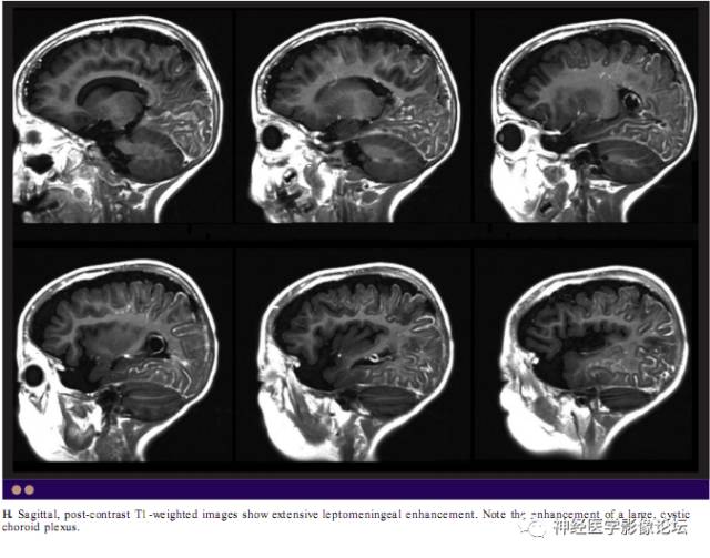
Leptomeningeal enhancement in patients with Sturge Weber syndrome is probably caused due to enhancement of the leptomeningeal angiomatosis(血管瘤病) and may allow early diagnosis of Sturge Weber syndrome. Enhancement of an enlarged ipsilateral choroid plexus(脉络丛) may be seen.
SYPHILIS
(梅毒)
Pathologically, meningovascular(脑膜血管) syphilis is manifested by widespread thickening of the meninges, with lymphocytic infiltration involving the meninges and around the small vessels. Contrast-enhanced MRI shows curvilinear, nonhomogeneous(不均匀) enhancement of the leptomeninges.