History: A 19-year-old man presents with progressive, right-sided hip pain.
病史:19岁男性,进行性右髋部疼痛。
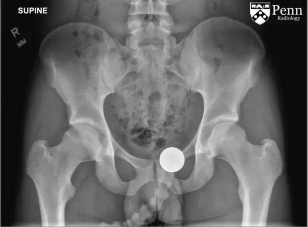
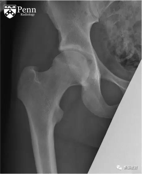
Frontal radiographs of the pelvis and right hip are shown below.
骨盆及右髋正位片如下。
CT images
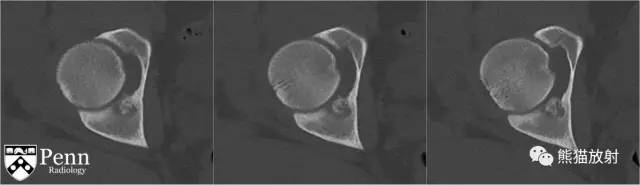
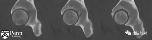
Unenhanced axial and sagittal CT images are shown below.
轴位、矢状位CT平扫图像如下。
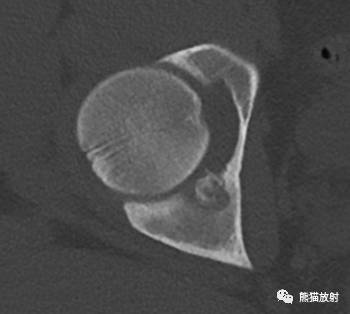
Findings
- Radiographs: There is a small, round, radiolucent lesion of the right acetabulum with a central focus of increased density.
- CT: The radiolucent lesion is seen in the cortex of the right acetabulum with a central sclerotic nidus.
影像表现:
平片:右侧髋臼可见一小类圆形透亮灶,其中心可见高密度灶。
CT:右侧髋臼骨皮质可见一低密度灶,并中心见硬化性瘤巢。
Differential diagnosis
- Osteoid osteoma
- Osteoblastoma
- Chronic cortical osteomyelitis
- Osteosarcoma
- Ewing’s sarcoma
鉴别诊断:
- 骨样骨瘤
- 骨母细胞瘤
- 慢性骨髓炎
- 骨肉瘤
- 尤文氏肉瘤
Diagnosis: Osteoid osteoma
诊断:骨样骨瘤
Key points 要点
Osteoid osteoma 骨样骨瘤
Pathophysiology
- Osteoid osteoma is a solitary, benign bone lesion, usually occurring in a long bone cortex.
- Its pathogenesis is controversial. Some believe it arises on an inflammatory basis. Some suggest it is an unusual healing and reparative process.
- Characteristically and unlike other bone tumors, it is composed of a nidus with a relatively immature central area and more mature calcified peripheral bone.
- It has little to no growth potential. They rarely exceed 1.5 cm.
病理生理学
- 骨样骨瘤是一种孤立性良性骨病,常见于长骨骨皮质。
- 发病机制不确定。部分人认为其起源于炎性基础;部分人观点认为其是一种不常见的愈合恢复过程。
- 病变与其它骨肿瘤不同,有一定特征性,由含有相对不成熟的中心区的瘤巢及更加成熟的钙化性的周围骨组成。
- 病变增长潜力非常小,大小很少超过1.5cm。
Epidemiology
- Osteoid osteoma is usually found in the lower extremities of children and young adults, with the proximal femur as the most common location.
- Most commonly occurs in the second and third decades.
- It is the third most common benign bone tumor.
- 10% to 11% of benign bone tumors
- 3% of all primary bone tumors
- The male-to-female ratio is 3:1.
流行病学
- 骨样骨瘤常见于儿童及青年下肢,最常见发病部位为股骨上端。
- 多见于二三十岁青年患者;
- 为第三位常见良性骨肿瘤;
- 占良性骨肿瘤的10%~11%;
- 占所有原发性骨肿瘤的3%;
- 男女发病率比例为3:1。
Clinical presentation
Most patients present with severe pain.
- The pain is characteristically increased at night.
- NSAIDs are used for pain relief.
- Prostaglandin production is believed to be a major factor.
- Duration of pain varies from weeks to years.
Osteoid osteomas are often misdiagnosed clinically.
临床表现
大部分患者表现为剧烈疼痛:
- 夜间疼痛;
- 服用NSAIDs使疼痛缓解;
- 前列腺素的分泌被认为是主要因素;
- 疼痛时间数周至数年。
骨样骨瘤在临床上常被误诊。
Imaging features
Radiographs:
- Classically, radiographs show an area of cortical thickening and sclerosis containing a lucent central nidus (< 1 cm).
- A nidus is present in 85% of cases.
- 20% intramedullary with less surrounding sclerosis.
- Subperiosteal location often has limited surrounding sclerotic response.
CT:
Commonly considered to be the most valuable imaging modality for diagnosis and localization.CT offers better visualization of the nidus.
MRI: Cortically based lesions are less conspicuous on MRI due to decreased spatial resolution and volume averaging.
Bone scan — technetium-99
- Useful if osteoid osteoma is suspected but not obvious on radiographs.
- Shows avid radiotracer uptake during vascular, blood-pool, and delayed phases.
- Classic “double-density” appearance is very specific.
Differential diagnoses
- Osteoblastoma — some believe it is the same lesion at a different stage of development
- Chronic cortical osteomyelitis
- Osteosarcoma
- Ewing’s sarcoma
影像表现
平片:
- 典型表现为骨皮质增厚、硬化,包含一透亮的瘤巢(<1cm);
- 85%的病变可见瘤巢;
- 20%的病变位于髓内,少有周围硬化;
- 骨膜下位置常见限制性的周围硬化反应。
CT:
常被认为是最有价值的影像诊断及定位的检查方法。CT能最好的显示瘤巢。
MRI:
由于较低的空间分辨率及部分容积效应,骨皮质的病变在MRI上多不显著。
骨扫描:
- 在平片上表现不明显但仍怀疑骨样骨瘤时较有用;
- 表现为明显摄取,由于血管、血池及延迟灌注;
- 典型的双密度表现非常特殊。
鉴别诊断:
- 骨母细胞瘤(一些人认为它与骨样骨瘤为同一病变的不同发展时期)
- 慢性骨髓炎
- 骨肉瘤
- 尤文氏肉瘤
Treatment
Conservative treatment:
- NSAIDs are used for pain relief.
- Osteoid osteomas may undergo spontaneous regression after several years.
Percutaneous — many options are available, including the following:
- MRI-guided cryotherapy
- CT-guided drilling of the nidus with ethanol injection
- Radiofrequency thermocoagulation
Surgery:
- Osteoid osteomas are often difficult to identify.
- Incomplete resection may lead to recurrence.
治疗
保守治疗:
- 服用NSAIDs缓解疼痛
- 可在几年后自然消退
介入治疗(包括几种有效的方法):
- MRI引导下冷冻治疗
- CT引导下向瘤巢内注入无水酒精
- 射频热凝术
手术治疗:
- 骨样骨瘤常很难确定;
- 切除不完全可导致复发。