第四脑室平底征(flat floor of fourth ventriclesign)
【影像学表现】
第四脑室平底征为头颅MRI上,脑干弥漫性肿胀,向后压迫第四脑室,使其前缘变平。
【临床意义】
第四脑室平底征见于脑干弥漫性星形细胞瘤,后者是儿童脑干胶质瘤中最常见的类型。弥漫性星形细胞瘤易向其低分化类型转变,即转化为间变型星形细胞瘤、甚至胶质母细胞瘤。脑干弥漫性星形细胞瘤根据其生长方式可分为外生型、播散型和内生型,以后者最为常见,肿瘤呈局限膨胀性生长。MRI显示脑干弥漫性增粗或局限型膨大,呈T1WI略低信号、T2WI高信号,增强后往往不强化。肿瘤占位效应较明显,可见邻近第四脑室受压,呈平底样改变。
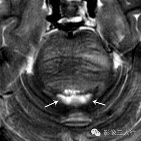
图1:头颅MRI提示脑干弥漫性肿胀、 向后压迫第四脑室,使其前缘变平(白箭)。
患者,女性36岁下巴肿胀,麻木不适


最后诊断:脑干胶质瘤
病理与临床特点
脑干弥漫性胶质瘤( diffuse brain stem gliomas)很少见于成人,因此也称儿童脑干胶质瘤,约占颅内肿瘤 2%及儿童脑肿瘤的 10%-15%,儿童病人脑干弥漫性胶质瘤仅次于小脑半球星形细胞瘤及髓母细胞瘤而居第3 位。2016年 WHO 分类中归属于弥漫性胶质瘤中的弥漫性中线胶质瘤( diffuse midline glioma),后者还包括弥漫性侵犯丘脑与脊髓的胶质瘤。本病通常为WHOⅡ级弥漫性星形细胞瘤,也可为毛细胞性星形细胞瘤,约 50%可向间变性星形细胞瘤或胶质母细胞瘤转化。偶见室管膜瘤及节细胞胶质瘤。可单独位于脑干局部,也可外向性生长,甚至脑脊液种植。位于顶盖及外向性生长的脑干胶质瘤预后较好。少数病例合并I 型神经纤维瘤病。肿瘤呈浸润性生长而不破坏脑干结构,因此多在晚期才出现临床症状。本病多见于儿童(约占 80%),发病高峰年龄为5 岁左右。临床表现与脑干神经核团受累及脑积水有关,如面部麻木、头晕、听力下降、头痛及呕吐。
Diffuse brain stem gliomas (diffuse brain stem gliomas) are rarely seen in adults, and are also referred to as pediatric brain stem gliomas, accounting for about 2% of intracranial tumors and 10%-15% of pediatric brain tumors. Diffuse brain stem gliomas in pediatric patients are the 3rd place only after astrocytoma and medulloblastoma in cerebellar hemisphere. Diffuse midline glioma (diffuse glioma) classified by WHO in 2016 also includes diffuse gliomas invading thalamus and spinal cord. The disease is usually Ⅱ diffuse astrocytoma, WHO can also be used for nonpilocytic astrocytoma, about 50% to anaplastic astrocytoma and glioblastoma. Ependymoma and ganglion glioma were found occasionally. It may be located locally in the brainstem alone, may also be extroverted growth, or even cerebrospinal fluid implantation. Brain stem gliomas located in the apex and extroversion have a better prognosis. A few cases were associated with neurofibromatosis type I. The tumor is invasive and does not destroy the brainstem structure, so the clinical symptoms usually appear in the late stage. The disease is more common in children (about 80%), the peak age of onset is about 5 years old. Clinical manifestations are associated with involvement of brainstem nuclei and hydrocephalus, such as facial numbness, dizziness, hearing loss, headache, and vomiting.
CT与 MRI 特点
①部位及形态:可发生于中脑、脑桥及延髓,脑桥最常见,向背侧外生性者可侵入小脑延髓池。肿瘤呈类圆形及膨胀性生长,邻近脑实质及脑池、第四脑室受压,向前外生性生长、包绕基底动脉。近 1/4 病例出现幕上梗阻性脑积水;
(1)Location and morphology: can occur in the middle brain, pons and medulla oblongata, pons is the most common, dorsal exophyte can invade the cerebellar medulla oblongata cisterna. The tumor showed a kind of round and expansive growth, adjacent to the brain parenchyma, cium and fourth ventricle compression, forward exogenous growth, around the basilar artery. Supratentorial obstructive hydrocephalus was found in nearly 1/4 cases.
②CT 由于颅底骨质的伪影,对脑干胶质瘤显示欠佳。平扫表现为低密度、等密度或混杂密度(约各占 1/3),仅约 5%可见代表少量钙化或出血的高或稍高密度;
(2) CT is not good for brain stem glioma due to the artifact of skull base. Plain scan showed low density, equal density or mixed density (about 1/3 each), and only about 5% showed high or slightly high density representing a small amount of calcification or bleeding.
③MR 对本病高度敏感。T1多为低信号,T2及 FLAIR 呈高信号。合并出血时表现为 T1及 T2 高信号,SW及 T2W 呈低信号。囊性变时呈T 低信号及 T2 高信号 FLAIR 低信号。位于顶盖时,表现为局部结节状肿块、导水管受压及第三脑室以上扩大,根据影像学表现即可做出诊断。中脑胶质瘤可向上、下分别延伸至丘脑与小脑,延髓胶质瘤还可向颈段脊髓扩展。增强扫描仅约 1/4 病人可见斑片状或环状强化。低级别者也可见较明显强化,这一点与幕上者不同;
(3)MR is highly sensitive to the disease. T1 mostly has low signal,T2 and FLAIR show high signal. High signal of T1 and T2, low signal of SW and T2W. There is T low signal and T2 hyperintensity FLAIR low signal in cystic degeneration. At the top of the lid, local nodular mass, compression of the aqueduct and enlargement of the third ventricle can be diagnosed according to imaging findings. Midbrain glioma can extend to thalamus and cerebellum respectively, and medullary medullary glioma can also extend to cervical spinal cord. Patchy or annular enhancement was seen in only about 1 in 4 patients on enhanced scan. The lower level also can see the obvious enhancement, this point is different from the screen;
④需注意的是,尽管脑干胶质瘤病理学为良性或低度恶性,随着时间的推移,肿瘤可逐渐増大与级别増髙,CT与MRⅠ显示脑干肿块体积増大、强化逐渐明显及范围扩大。毛细胞星形细胞瘤预后最佳,其影像学特点囊实性病变,实性部分明显强化。
(4) is important to note that although the brainstem glioma pathology of benign or low-grade malignant, with the passage of time, the level of tumor can be gradually raised large and raised high, CT and MR Ⅰ shows brainstem tumor volume rights, strengthening the increasingly obvious and expanded. Hair cell astrocytoma has the best prognosis. Its imaging features are cystic and solid lesions with obvious enhancement of solid parts.
鉴别诊断
①脑干脑炎,起病急,多有前驱感染,影像学上无基底动脉包绕征象;
(1) brainstem encephalitis, the onset of acute, more prodromal infection, imaging no basilar artery wrapped signs;
②多发性硬化,临床表现为多期相特征,影像学可见脑白质多发病变;
(2) multiple sclerosis, clinical manifestations of multiphase features, imaging can see multiple lesions of white matter;
③海绵状血管瘤,脑干胶质瘤CT 平扫为高密度时需与海绵状血管瘤后者 MRI 上可见典型的爆米花状混杂信号及 T2W及 T2WI或 SWI 低信号环;
(3) cavernous hemangioma, brainstem glioma CT plain scan for high density and cavernous hemangioma, the latter MRI can see the typical popcorn like mixed signals and T2W and T2WI or SWI low signal ring;
④脑干脓肿,临床有感染症状,增强扫描呈环状强化及水肿,治疗后病变减小;
(4) brainstem abscess, clinical infection symptoms, enhanced scan showed annular enhancement and edema, reduced lesions after treatment;
⑤渗透压脑病,有代谢紊乱及快速纠正低钠史。
(5)Osmotic encephalopathy, metabolic disorders and rapid correction of low sodium history.
简要讨论
脑干胶质瘤特点为脑干局部膨大,MRI 显示最佳,T2及 FLAIR 为高信号,可包绕基底动脉及延伸至小脑延髓池,部分病例可见强化。
Cerebral stem glioma is characterized by local enlargement of the brainstem,MRI shows best,T2 and FLAIR are hyperintensity, and can wrap around the basilar artery and extend to the cerebellar medullary cistern, with enhancement seen in some cases.