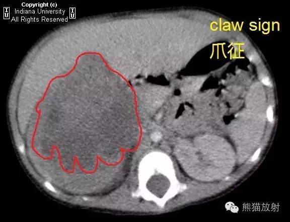
History: A 2-year-old child presents with nausea and vomiting.
病史:2岁儿童,恶心、呕吐。
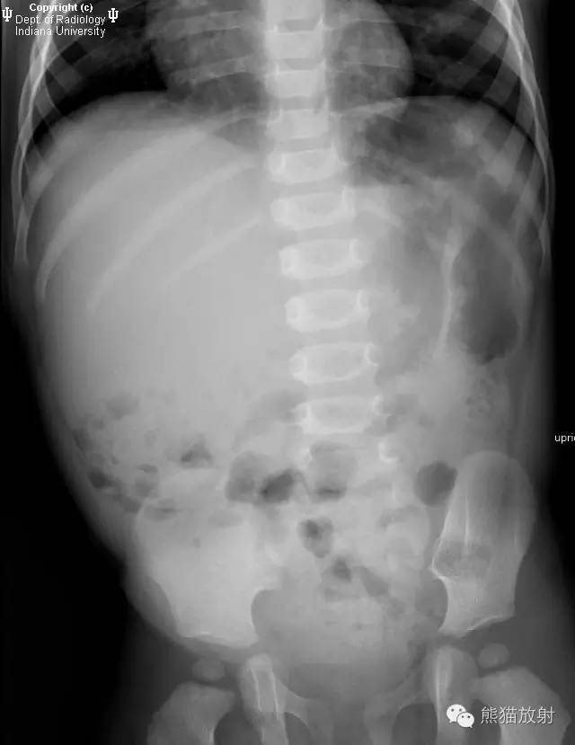
Abdominal radiograph is shown below.
腹部平片如下所示。
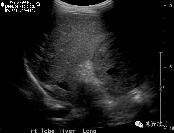
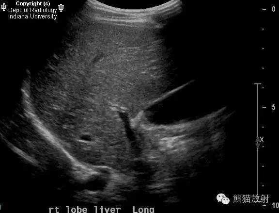
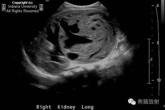
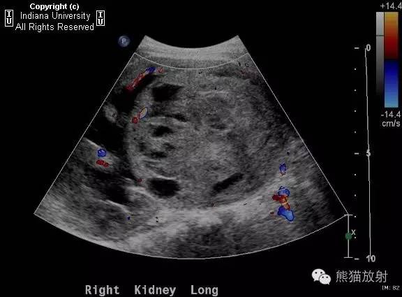
Ultrasound images are shown below.
超声图像如下所示。
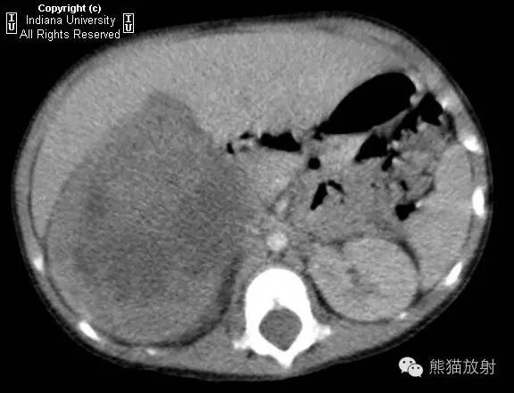
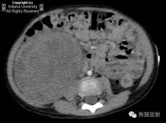
CT images are shown below.
CT图像如下所示。
Findings
There is a mixed echogenic mass in the right kidney containing solid and cystic components. CT confirms a large mass centered at the lower pole of the right kidney.
影像表现:
右肾见一混合回声肿物,含实性和囊性成分。CT证实以右肾下极为中心见一巨大肿物。
Differential diagnosis:
Wilms\’ tumor
Neuroblastoma
Renal cell carcinoma
鉴别诊断:
Wilms瘤
神经母细胞瘤
肾细胞癌
Discussion
Wilms\’ tumor is the most common malignancy of the kidneys in children. The tumor can grow asymptomatically and is often identified on physical exam as an abdominal mass after significant growth. Children are usually identified with the tumor between ages 1 year and 5 years. Wilms\’ tumor occurs most commonly in other wise normal children, but there is an association with Beckwith-Wiedemann syndrome, congenital hemihypertrophy, trisomy 18, and sporadic aniridia.
Wilms\’ is a tumor consisting of primitive metanephric blastemal elements. Prognosis is very good with a cure rate of greater than 90%. Nephrectomy is often performed with chemotherapy used for unresectable tumors.
讨论
- Wilms瘤是小儿最常见的肾脏恶性肿瘤;
- 无明显临床症状,常因肿瘤体积明显增大后发现;
- 多见于1-5岁;
- 可与下列疾病相关:伯-韦综合征、先天性半侧肥大、18三体综合征、散发性先天性无虹膜;
- Wilms瘤包含原始后肾胚细胞元素;
- 预后较好,治愈率超过90%;
- 治疗采取肾切除术,不能切除的采用化疗。
Radiologic overview
Given that Wilms\’ affects the pediatric population, the initial imaging study is usually ultrasound. On ultrasound, Wilms\’ appears as a large, heterogeneous mass of the kidney. Wilms\’ often invades the renal vein and inferior vena cava (IVC), and there may be signs of tumor thrombus invasion on color Doppler.
After identification on ultrasound, chest CT is usually the next study to stage the tumor. The lungs are the most common location for metastasis. On CT, there is a large heterogeneous mass that replaces the kidney and displaces adjacent organs. There is no engulfment of vessels as seen with neuroblastoma and a “claw sign” is often seen as the tumor displaces the normal renal parenchyma. In 5% of cases, Wilms\’ involves the contralateral kidney, so special attention should be made in evaluating both kidneys.
In this case, there is a heterogenous mass involving the right kidney. On CT, the claw sign is seen as the mass displaces the normal renal parenchyma. The patient underwent surgical resection.
影像表现
- 患者主要为儿童,首要检查方法为超声;表现为肾脏体积巨大不均质回声肿物。
- 常侵犯肾静脉及下腔静脉,彩色多普勒超声可见癌栓征象。
- 超声检查后,下一步行胸部CT进行肿瘤分期,肺是最常见的转移部位。
- CT:肾脏被巨大不均质肿物所替代,周围脏器受压移位。肿瘤边缘呈“爪征”替代正常肾实质,不会像神经母细胞瘤那样侵犯血管。5%的患者对侧肾脏可受累。
- 本例,右肾可见一不均质肿物,边缘呈爪样替代正常肾实质。肿瘤被手术切除。
Key points
- Wilms\’ is the most common renal tumor in children. Peak age of incidence is 3 years.
- Wilms\’ can grow to a very large size before it is detectable on exam or is symptomatic.
- Ultrasound is the usual initial imaging test and will show a large, heterogenous mass arising from the kidney.
- There can be invasion of the renal vein and IVC.
- Wilms\’ tends to displace vessels rather than envelop them, which is what differentiates it from neuroblastoma.
知识点
- Wilms瘤是儿童最常见的肾脏肿瘤,发病年龄高峰为3岁;
- 临床无明显症状,多在生长至很大体积后检查发现;
- 超声是常用的首要检查方法,表现为肾脏巨大回声不均的肿物;
- 肿瘤可侵犯肾静脉及下腔静脉;
- Wilms瘤一般使周围血管移位而非包埋,此征象可与神经母细胞瘤相鉴别。