History and CT images
A 30-year-old man with chest pain and shortness of breath.
男,30岁,胸痛,呼吸困难。
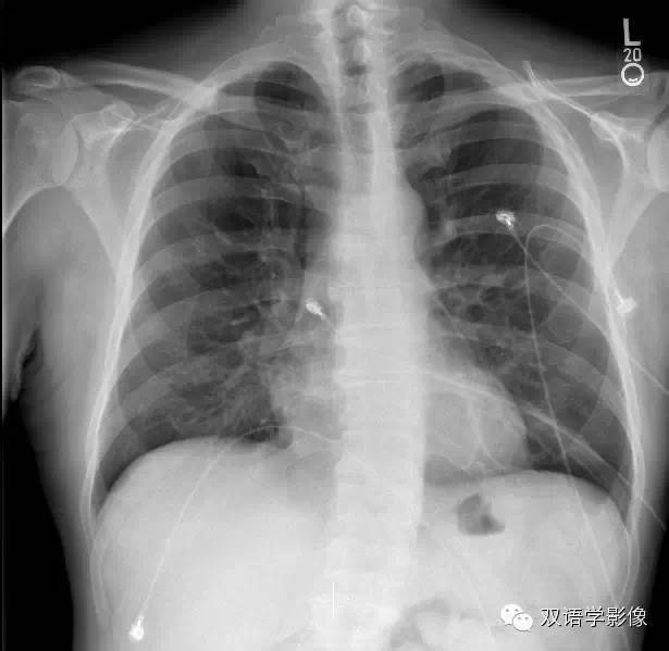
1.Alveolar consolidation(肺实变) is present.
True
False
2.Cardiomegaly (心影增大)is present.
True
False
3.Mediastinal adenopathy(纵膈淋巴结肿大) is present.
True
False
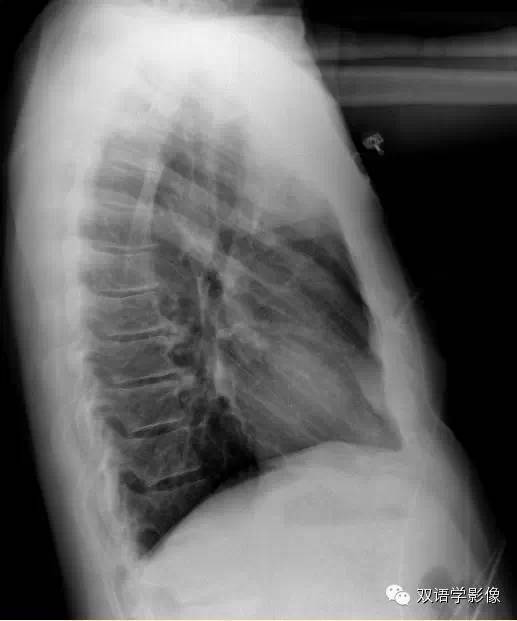
4.There is a tracheal abnormality(气道异常).
True
False
(请做出您的选择,答案见本文后方)
CT of the chest, PE protocol
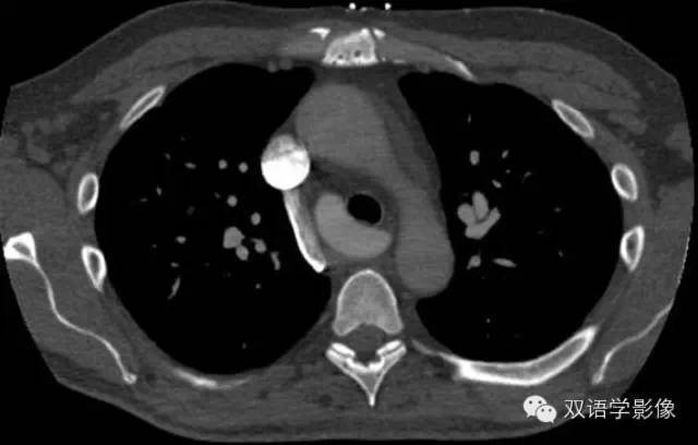

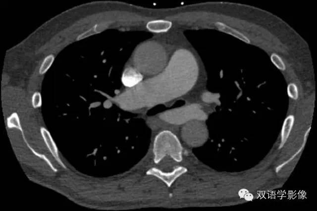
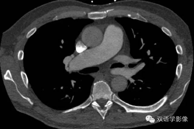
5.What is the etiology of the tracheal narrowing?
Aberrant vessel
Adenopathy
Esophageal narrowing
Neurogenic tumor
Pulmonary embolism
6.Which vessel is aberrant?
Left inferior pulmonary vein
Left pulmonary artery
Left subclavian artery
Left superior pulmonary vein
Right pulmonary artery
正确答案
- False
- False
- False
- True
There is indentation of the posterior trachea seen on the lateral film.
侧位像可见气管后方压迹。
- Aberrant vessel
There is aberrant course of one of the pictured vessels.
- Left pulmonary artery
The left pulmonary artery arises from the right pulmonary artery and courses between the trachea and esophagus
左肺动脉起源于右肺动脉,走行于气管与食管之间。
Findings and diagnosis
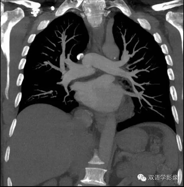
Findings
- PA and lateral chest x-ray: Indentation on the posterior aspect of the trachea on the lateral view.
X线胸部正侧位片:侧位像可见气管后方压迹。
- Contrast-enhanced CT with attention to the pulmonary arteries: Aberrant left pulmonary artery arising from the right pulmonary artery with narrowing of the adjacent trachea and left mainstem bronchus, as well as mass effect on the anterior esophagus.
CT增强扫描示左肺动脉起源于右肺动脉,临近主气管及左侧主支气管狭窄,食管受压。
Differential diagnosis
- Pulmonary artery sling 肺动脉吊带
- Bronchogenic/foregut cyst 支气管/前肠囊肿
- Tracheobronchomalacia 气管支气管软化
- Bronchial atresia 支气管闭锁
Diagnosis:
Type I pulmonary artery sling
肺动脉吊带I型